Cell Division, Reproduction & Development — Print · Life Science: Biology, Grade 10
Connect chromosome continuity, variation, regulated growth, and specialization.
Printable chapter lessons and review.
Mitosis & the Cell Cycle
Learning goals
- I can describe the stages of the cell cycle and the four phases of mitosis.
- I can explain how mitosis produces genetically identical cells for growth, repair, and asexual reproduction.
Readings for this unit
Before this unit
Identify chromosomes and distinguish replication from division.
Use three flexible study sessions: core explanation and Parts 1–2; Parts 3–4 with their reasoning checks and models; then the whole-unit application, worked examples, independent practice, and exit check. Allow about 50–60 minutes per session, including diagrams, data work, and notebook explanations. Attempt each check before opening its guidance. Schedule chapter investigations, review, and exams separately; add time where prerequisites are uncertain. Trace the explained visual parts and write a first model, then attempt the illustrated and changed independent cases. In the third session explain a correction and revise the opening account using evidence.
Start with something to explain
Why does DNA replication not make an ordinary cell’s daughters twice as diploid?
Illustrative evidence case. A copied chromosome has two joined sister chromatids before their separation; nuclear division and cytokinesis have distinct roles.
Notice and wonder
Record two observations using the stated quantities or relationships. Keep observations separate from possible causes. Write one question that a new measurement could help answer.
Why does DNA replication not make an ordinary cell’s daughters twice as diploid?
Your first model
Label chromosome sets, centromeres and DNA copies at each stage.
Build the language from the idea
- cell cycle
- The full, repeating sequence of growth, DNA copying, and division that a cell passes through.
- interphase
- The long part of the cell cycle before division, when a cell grows, does its work, and copies its DNA.
- mitosis
- Division of the nucleus that produces two genetically identical nuclei.
- sister chromatids
- The two identical copies of a chromosome, joined together, that result when a cell copies its DNA.
- cytokinesis
- The final step of division, in which the cytoplasm splits to form two separate daughter cells.
- daughter cell
- One of the two new cells produced when a parent cell divides.
- centromere
- A chromosome region associated with chromatid organization and attachment machinery; joined sister units are counted as one chromosome before separation.
- G₁, S and G₂
- Typical interphase stages: growth/preparation before copying, DNA synthesis, and preparation after copying.
A single fertilized egg becomes a body of tens of trillions of cells, a cut knits itself closed, and worn-out blood and skin cells are replaced by the billions every day. All of that depends on one carefully controlled process: cell division. But a cell cannot simply split in half whenever it likes — it moves through an orderly, repeating sequence called the cell cycle, and dividing is only the final act.
Most of a cell's life is spent in interphase, the long 'everyday' stretch before division. During interphase the cell grows, carries out its normal work, and — critically — copies its nuclear DNA in S phase, so that every chromosome is duplicated. After that copying, each chromosome consists of two identical halves called sister chromatids, joined together at a pinched-in point. Duplicating first is the whole trick: just as you would photocopy every page of a manual before splitting it into two complete sets, copying the DNA in advance normally allows both future cells to receive a full set.
Then comes mitosis, the division of the nucleus, which unfolds in four smooth phases. In prophase, the duplicated chromosomes coil up tight and become visible, and the nuclear envelope breaks down. In metaphase, the chromosomes line up single file across the middle of the cell. In anaphase, the sister chromatids are pulled apart, one of each pair dragged toward opposite ends of the cell. And in telophase, a new nuclear envelope forms around each of the two separated sets. A handy way to remember the order is PMAT: Prophase, Metaphase, Anaphase, Telophase.
Mitosis divides the nucleus, but one step remains: cytokinesis, in which the rest of the cell pinches in two, splitting the cytoplasm so that each new nucleus ends up in its own separate cell. The outcome is two daughter cells — and because each one received a complete, identical set of chromosomes copied from the same original, the two normally inherit corresponding chromosome sets; copying or segregation errors can introduce differences. That fidelity is the entire point of mitosis.
This is why a scraped knee heals with matching skin rather than random tissue, why you grow, and why single-celled organisms such as amoebas can reproduce entirely on their own — some eukaryotic asexual reproduction uses mitosis; bacteria divide through other mechanisms. Precisely because division is so powerful, the cell cycle is tightly regulated by molecular 'checkpoints' that halt the process if something is wrong. When the genes controlling those checkpoints are damaged by mutation, cells can begin dividing uncontrollably, ignoring the normal stop signals — and that runaway division is the root of cancer.
DNA replication occurs before the division phase of a typical cell cycle. Mitosis separates duplicated chromosome sets, while cytokinesis divides the cell. These related events should not be treated as the same process.
In a diploid somatic-cell model, mitosis generally preserves chromosome number and produces genetically similar daughter cells. Replication makes sister chromatids but does not double the count of chromosome centromeres. State the counting convention when discussing chromosomes.
Cell-cycle checkpoints coordinate growth, DNA integrity and division. A repair or arrest response can limit propagation of damage. A diagram of phases represents a regulated cycle with variable timing, not a clock in which every cell spends equal time in every stage.
Build your understanding
Use three flexible study sessions: core explanation and Parts 1–2; Parts 3–4 with their reasoning checks and models; then the whole-unit application, worked examples, independent practice, and exit check. Allow about 50–60 minutes per session, including diagrams, data work, and notebook explanations. Attempt each check before opening its guidance. Schedule chapter investigations, review, and exams separately; add time where prerequisites are uncertain.
Part 1 · The cycle is mostly interphase
Interphase includes G₁, DNA synthesis in S phase, and preparation in G₂; cells can also enter other states. A replicated chromosome has two sister chromatids, initially associated at a centromere region. In the usual counting convention, it remains one chromosome until its sisters separate. Ploidy counts chromosome sets, not DNA amount. Therefore a diploid cell can double its DNA before division while remaining diploid. State which quantity is being counted rather than treating every X-shaped arm as a chromosome.
Read the visual model
Identify the labeled structures and units, then trace the arrows or compare values under the caption’s stated conditions. Separate the displayed evidence from a further inference. Write your reasoning before opening the guidance.
A cell has 8 chromosomes before S phase. What are its chromosome and chromatid counts just after replication?
Visual reasoning — after your attempt
It has 8 replicated chromosomes with 16 chromatids under the stated centromere convention; its chromosome-set count has not doubled.
The cell cycle divides into interphase and division. Interphase has three parts: in G1 the cell grows and builds proteins and organelles, in S it replicates its entire DNA so that every chromosome becomes two attached copies, and in G2 it completes preparations and assembles the structures that will move the chromosomes. Mitosis and cytokinesis then separate the copies and split the cell.
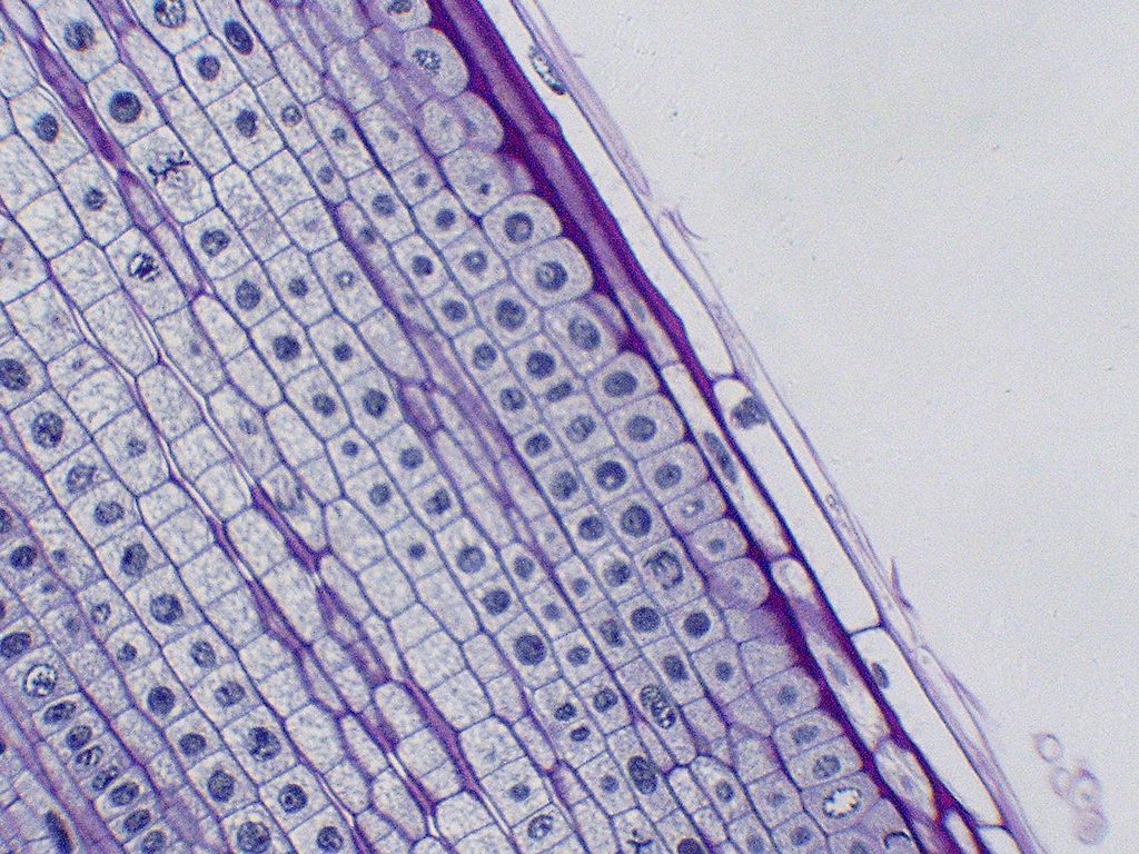
Proportion matters here. In a typical dividing cell, interphase occupies the large majority of the cycle and mitosis a small fraction, which is why most cells in a microscope field show no visible chromosomes. Calling interphase a resting stage inverts the truth: it is when replication and most synthesis occur, and it only looks quiet because light microscopy cannot resolve those activities.
Pause and explain
Why do most cells in a root-tip field show no visible chromosomes?
Check your reasoning after answering
Most cells are in interphase, which occupies the majority of the cycle, and during interphase the chromosomes are not condensed enough to be visible as distinct structures under a light microscope.
Part 2 · Four phases that divide duplicated chromosomes
During mitosis, duplicated chromosomes condense, attach to spindle structures, align and separate their sisters toward opposite poles before new nuclei organize. After sister separation, each former chromatid is counted as a chromosome in its destination set. Cytokinesis divides cellular contents and boundary, a related but distinct process. The usual result preserves chromosome number in daughter nuclei. Mutations, segregation errors and cell-state differences mean “identical” should be treated as an ideal copying outcome rather than an exceptionless guarantee.
Read the visual model
Identify the labeled structures and units, then trace the arrows or compare values under the caption’s stated conditions. Separate the displayed evidence from a further inference. Write your reasoning before opening the guidance.
What is separated in typical mitosis: homologous pairs or sister chromatids?
Visual reasoning — after your attempt
Sister chromatids separate; homolog pairing and reduction belong to the meiosis-I comparison.
Mitosis is conventionally divided into four phases. In prophase the duplicated chromosomes condense and become visible, and the nuclear envelope breaks down. In metaphase they align along the cell's midline, attached to fibres from both poles. In anaphase the attached copies separate and move to opposite poles. In telophase the chromosomes decondense and nuclear envelopes re-form, and cytokinesis divides the cytoplasm.
The names are less important than the logic: every step exists to ensure that each daughter receives exactly one copy of each chromosome. Alignment before separation is what makes equal division possible, and attachment to fibres from both poles is what ensures the two copies travel in opposite directions. A fault in attachment produces daughter cells with wrong chromosome numbers.
Pause and explain
Why must chromosomes align at the midline and attach to both poles before anaphase begins?
Check your reasoning after answering
Alignment with attachments running to opposite poles is what guarantees the two copies of each chromosome are pulled apart in opposite directions. Without it, both copies could travel to the same daughter cell, leaving the other short.
Part 3 · Checkpoints, and what their failure means
A checkpoint is a regulatory interaction that coordinates progression with conditions such as DNA integrity and spindle attachment. Repair, arrest and other responses can limit the propagation of damage. Cancer involves altered growth regulation and additional changes in cellular behavior; a single missed checkpoint is not a complete diagnosis or explanation of every tumor. Rate and timing differ among tissues. A cell-cycle diagram should show controlled transitions and distinguish damaged DNA, attempted repair and the later fate of the cell.
Read the visual model
Identify the labeled structures and units, then trace the arrows or compare values under the caption’s stated conditions. Separate the displayed evidence from a further inference. Write your reasoning before opening the guidance.
Does the mere observation of cell division establish failed regulation?
Visual reasoning — after your attempt
No. Division is also part of normal growth and renewal; interpretation needs the conditions, tissue behavior and regulatory evidence.
The cycle is regulated at checkpoints where the cell verifies conditions before proceeding: whether it is large enough and has adequate resources, whether DNA replication is complete and undamaged, and whether chromosomes are properly attached before separation. If a check fails, the cycle halts for repair, or the cell is destroyed rather than allowed to divide.
Cancer is a loss of this regulation. Cells with damaged control genes divide without regard to the organism's needs or to the state of their own DNA, so faults accumulate and a mass forms that consumes resources and can invade surrounding tissue. The abnormality is in the control of division, not in division itself, which is why tissues that normally divide rapidly, such as bone marrow and intestinal lining, are not themselves abnormal.
Pause and explain
Bone-marrow cells divide very rapidly and are not cancerous. What distinguishes rapid normal division from cancerous division?
Check your reasoning after answering
Normal rapid division remains regulated: it responds to the organism's needs and halts when checkpoints detect damage or incomplete replication. Cancerous division proceeds regardless of those signals, so rate alone does not distinguish the two — the presence or absence of control does.
Part 4 · Estimating phase durations from counts
An asynchronous sample contains cells at different stages. If cells cycle comparably and the population is suitably sampled, a stage’s fraction can approximate its fraction of cycle time. Thirty prophase cells among 200 gives 15%; a specified 24-hour cycle then gives 3.6 hours. Different division rates, noncycling cells, cell death, sampling and classification errors can break the inference. A snapshot is not direct timing of one cell, and equal diagram panels do not mean equal stage durations.
Read the visual model
Identify the labeled structures and units, then trace the arrows or compare values under the caption’s stated conditions. Separate the displayed evidence from a further inference. Write your reasoning before opening the guidance.
Why is the 3.6-hour value a conditional estimate rather than an observed stopwatch measurement?
Visual reasoning — after your attempt
It converts a population fraction using stated comparable-cycle assumptions; a time-lapse of individual cells measures duration differently.
Phase durations can be estimated without watching a single cell through its cycle. In a tissue whose cells divide asynchronously, the proportion of cells caught in a phase approximates the proportion of the cycle that phase occupies. If 15 percent of cells are in mitosis and the cycle takes 20 hours, mitosis takes about 3 hours.
The method rests on assumptions worth stating. Cells must be dividing independently rather than in step, the sample must be representative of the tissue, and fixation must not favour one phase. Phases are also unequal in how easily they are recognized — metaphase is distinctive, early prophase less so — which biases counts toward the phases that are easy to score.
Pause and explain
Why does this counting method fail in a tissue whose cells divide synchronously?
Check your reasoning after answering
If cells divide in step, the proportion in each phase at one instant reflects where the whole population happens to be rather than the relative lengths of the phases. A single snapshot could show nearly all cells in one phase even if that phase is brief.
Apply the whole unit · Estimate phase lengths in a root tip
Illustrative counts from a stained onion root-tip squash, 500 cells scored by one observer. Interphase 425; prophase 32; metaphase 18; anaphase 11; telophase 14. Independent evidence gives the full cycle length in this tissue as 20 hours.
Your task
Calculate the proportion of cells in each phase and the estimated duration of each in hours, state which phase is briefest and by how much it differs from the longest mitotic phase, and explain the three assumptions the estimate depends on. Then identify the most likely source of systematic error in these counts and how you would reduce it.
What your response should show
- Choose the appropriate biological model or relationship and state the conditions it assumes.
- Show the counts, rates, ratios, or intermediate reasoning the question requires.
- Use the supplied evidence to justify the conclusion and separate observation from inference.
- Identify a meaningful limitation, uncertainty, or next measurement.
Response guidance — after your attempt
Proportions: interphase 425/500 = 0.850; prophase 0.064; metaphase 0.036; anaphase 0.022; telophase 0.028.
Durations at 20 hours: interphase 17.0 h; prophase 1.28 h; metaphase 0.72 h; anaphase 0.44 h; telophase 0.56 h.
Compare: anaphase is the briefest at about 0.44 h, roughly a third of prophase's 1.28 h, and mitosis as a whole accounts for 3.0 of the 20 hours.
State the assumptions: cells divide asynchronously, the 500 cells are representative of the tissue, and fixation and staining do not preserve or reveal one phase preferentially.
Identify the error source: prophase and telophase are harder to score than metaphase, because condensing and decondensing chromosomes grade into interphase appearance, so counts of those phases are the least reliable.
Reduce it: have two observers score the same fields independently and compare, use a defined criterion for each phase, and count more cells across several root tips.
Interphase accounts for 85 percent of the cells and therefore about 17.0 of the 20 hours, with mitosis occupying the remaining 3.0 hours: prophase about 1.28 h, metaphase 0.72 h, anaphase 0.44 h, and telophase 0.56 h. Anaphase is the briefest, about a third the length of prophase, which fits its role as a rapid separation step. The estimate depends on cells dividing asynchronously, the sample being representative, and the preparation not favouring any phase. Its weakest point is phase identification at the boundaries: early prophase and late telophase resemble interphase, so those counts are the least reliable and errors there shift the estimates for both. Independent scoring by two observers with explicit criteria, applied to more cells from several root tips, would reduce that bias and allow the spread between observers to be reported alongside the estimate.
Scientific references and adapted source figures
Parent / teacher teaching notes
The phase-counting calculation is the most valuable twenty minutes in this unit, because it converts a memorized list into a measurement and it reinforces the sampling reasoning from the first chapter. Correct "interphase is when the cell rests" whenever it appears; the S phase is the single most consequential part of the cycle. Keep the cancer discussion on the regulation point — rapid division is normal in several healthy tissues — which is both accurate and avoids alarming conclusions drawn from division rate alone. Ask for an explanation of each label, arrow and axis before calculation. Keep first and revised models; attempt the changed case before consulting guidance. Distinguish invented instructional records from named historical sources. Source chromosome symbols are allele-category models, not DNA sequences. These independently authored high-school supplements are not the full OpenSciEd teacher editions or an endorsement.
An animated stepper showing the phases of mitosis, from a cell with copied chromosomes lining up in the middle to two identical daughter cells.
The key idea
In the standard cycle, S phase copies nuclear DNA before mitosis separates sister chromatids. Cytokinesis partitions the cell. Daughter nuclei normally retain corresponding chromosome sets; this is distinct from a guarantee of identical cells or error-free copying.
Cells After Mitosis
cells after n divisions = 2^n
one cell dividing by mitosis n times in a row, with no cell death.
Worked examples
Example 1
- Copying the DNA duplicates each chromosome into two sister chromatids joined together — it does not create separate new chromosomes.
- So the chromosome count stays 46, because two joined sister chromatids still count as one chromosome.
- But each of the 46 chromosomes now has 2 chromatids: 46 x 2 = 92 chromatids.
Example 2 · Worked application
- DNA replicates before division, forming sister chromatids.
- Mitosis separates sister chromatids into daughter nuclei.
- Each daughter retains the starting chromosome count under normal division.
Chromatid doubling after replication is not a doubling of chromosome count when counted by centromeres.
Example 3
- Each round of mitosis doubles the number of cells: 1, 2, 4, 8, 16. That is 2^4 = 16 cells.
- Mitosis preserves the full chromosome set at every division, so the number per cell never changes.
- Each of the 16 cells therefore has the same 46 chromosomes as the original.
Example 4
- Replication makes sister chromatids without changing the stated chromosome number.
- Normal separation gives each daughter the original set size.
Example 5 · Find and fix the mistake
- Test the claim against the stated conditions: Chromatid doubling after replication is not a doubling of chromosome count when counted by centromeres.
- Replication supplies copies that separate; normal mitosis preserves the count.
Chromatid doubling after replication is not a doubling of chromosome count when counted by centromeres.
Example 6 · Reason from the visual evidence
- A checkpoint is a regulatory interaction that coordinates progression with conditions such as DNA integrity and spindle attachment. Repair, arrest and other responses can limit the propagation of damage. Cancer involves altered growth regulation and additional changes in cellular behavior; a single missed checkpoint is not a complete diagnosis or explanation of every tumor. Rate and timing differ among tissues. A cell-cycle diagram should show controlled transitions and distinguish damaged DNA, attempted repair and the later fate of the cell.
- No. Division is also part of normal growth and renewal; interpretation needs the conditions, tissue behavior and regulatory evidence.
Original control-decision abstraction, not a cancer diagnostic model or exhaustive checkpoint network.
Avoid these mistakes
- DNA replication occurs before mitosis, not during every mitotic stage.
Check your understanding
A diploid cell has 12 chromosomes before replication. How many chromosomes does each daughter cell normally have after mitosis and cytokinesis?
Discuss the answer
Each daughter normally has 12 chromosomes in this model.
Why should mitosis and cytokinesis be named separately?
Discuss the answer
Mitosis distributes nuclear chromosome sets; cytokinesis partitions the remaining cell and boundary. They are coordinated but not the same event.
Return to the phenomenon
Return to the initial case. Why does DNA replication not make an ordinary cell’s daughters twice as diploid? Revise your drawing or explanation using two named ideas and one stated limitation.
Compare explanations after your attempt
Replication changes DNA amount, mitosis distributes sister copies and cytokinesis partitions the cell; counting conventions and regulation matter.
What should we investigate next?
Which comparison would test a remaining uncertainty? State what you would change, keep comparable and measure.
Investigate, explain and revise
Complete these tasks after the teaching parts. Preserve your first model and a supported revision; use guidance after a real attempt.
Activity 1 · Return to the opening case
Use the opening question, defined terms, Parts 1–4 and your notebook. The supplied-data route needs no equipment or external account.
Your task
Return to the initial case. Why does DNA replication not make an ordinary cell’s daughters twice as diploid? Revise your drawing or explanation using two named ideas and one stated limitation.
Success criteria
- Use relevant stated observations.
- Explain the mechanism with a labeled model.
- Distinguish quantities and assumptions.
- Describe a revision and a useful next test.
Hint
Follow objects, materials and information separately. Check what each measured quantity means.
Response guidance — discuss after your attempt
Replication changes DNA amount, mitosis distributes sister copies and cytokinesis partitions the cell; counting conventions and regulation matter.
Activity 2 · Transfer to a changed case
Cover the worked guidance and use the unit explanations/figures to solve a changed problem.
Your task
A diploid model starts with 10 chromosomes. Track chromosomes, chromatids and daughter-cell chromosome number through replication and normal mitosis/cytokinesis.
Success criteria
- Use the stated measurements or conditions, with correct units.
- Connect the result to the biological mechanism or evidence relationship.
- Identify the assumption, model limit or further evidence requested.
Hint
Name the stated conditions, select the relevant mechanism and show intermediate reasoning.
Response guidance — discuss after your attempt
After S phase there are 10 replicated chromosomes and 20 chromatids; during separation each former chromatid becomes a chromosome. Each daughter receives 10 chromosomes, preserving diploidy in the model.
Words to know
- cell cycle
- The full, repeating sequence of growth, DNA copying, and division that a cell passes through.
- interphase
- The long part of the cell cycle before division, when a cell grows, does its work, and copies its DNA.
- mitosis
- Division of the nucleus that produces two genetically identical nuclei.
- sister chromatids
- The two identical copies of a chromosome, joined together, that result when a cell copies its DNA.
- cytokinesis
- The final step of division, in which the cytoplasm splits to form two separate daughter cells.
- daughter cell
- One of the two new cells produced when a parent cell divides.
- centromere
- A chromosome region associated with chromatid organization and attachment machinery; joined sister units are counted as one chromosome before separation.
- G₁, S and G₂
- Typical interphase stages: growth/preparation before copying, DNA synthesis, and preparation after copying.
The cell cycle's checkpoints and its guardian
The cell cycle isn't a free-for-all; it pauses at checkpoints where the cell inspects its own work before continuing. A protein called p53, nicknamed 'the guardian of the genome,' halts the cycle when it detects damaged DNA, giving the cell time to repair the damage — or, if the damage is too severe, ordering the cell to self-destruct. Because p53 is so central to control, it is mutated in roughly half of all human cancers.
When mitosis loses its brakes
Cancer is, at its core, mitosis without proper control. Mutations in the genes that normally start and stop division can cause a cell to divide relentlessly, ignoring the signals that should tell it to pause or die. The descendants pile up into a tumor, and some may break away and spread. This is why many treatments — chemotherapy and radiation — are aimed at cells that are dividing rapidly, though that also explains their side effects.
Not all cells keep dividing
As an organism develops, most cells differentiate — they specialize into skin, muscle, nerve, and other types — and many then slow or stop dividing. Mature neurons and heart muscle cells almost never divide again, which is a major reason serious brain or heart damage is so hard to repair compared with a scraped knee. Stem cells, which keep the ability to divide and specialize, are the body's built-in repair kit and a major focus of modern medicine.
The Hayflick limit and 'immortal' cells
In the 1960s, Leonard Hayflick discovered that normal human cells divide only about 40 to 60 times before they stop for good — the Hayflick limit, tied to those shrinking telomeres. Cancer cells cheat this limit by switching on an enzyme, telomerase, that rebuilds telomeres, letting them divide without end. This 'immortality' is part of what makes cancer so persistent.
The cells that never stopped dividing
In 1951, cells taken from a cancer patient named Henrietta Lacks did something unusual: they kept dividing indefinitely in the lab. These 'HeLa' cells became the first immortal human cell line and have been used in research from the polio vaccine to cancer studies. Their story — the cells were taken without her knowledge or consent — has also become a landmark case in the ethics of medical consent and who controls a patient's biological samples.
In the real world
Because many cancer treatments target fast-dividing cells, they also strike other rapidly dividing tissues — hair follicles and the lining of the gut — which is why chemotherapy often causes hair loss and nausea.
Exit check
A cell has 12 chromosomes before replication. How many chromosomes and chromatids are present after copying, before separation?
Check your explanation after answering
There are 12 replicated chromosomes and 24 chromatids, counting chromosomes by centromeres while sister chromatids remain joined.
Explain your reasoning without notes. If a key idea is missing, revisit the lesson and try again.
Meiosis & Sexual Reproduction
Learning goals
- I can compare meiosis and mitosis and explain how meiosis produces haploid gametes.
- I can explain how crossing over, independent assortment, and fertilization create genetic variation.
Readings for this unit
Before this unit
Distinguish homologous chromosomes from sister chromatids.
Use three flexible study sessions: core explanation and Parts 1–2; Parts 3–4 with their reasoning checks and models; then the whole-unit application, worked examples, independent practice, and exit check. Allow about 50–60 minutes per session, including diagrams, data work, and notebook explanations. Attempt each check before opening its guidance. Schedule chapter investigations, review, and exams separately; add time where prerequisites are uncertain. Trace the explained visual parts and write a first model, then attempt the illustrated and changed independent cases. In the third session explain a correction and revise the opening account using evidence.
Start with something to explain
How can a haploid nucleus still contain duplicated chromosomes?
Illustrative evidence case. Set count falls in meiosis I before sisters separate in meiosis II.
Notice and wonder
Record two observations using the stated quantities or relationships. Keep observations separate from possible causes. Write one question that a new measurement could help answer.
How can a haploid nucleus still contain duplicated chromosomes?
Your first model
Label homologs, sisters and per-nucleus counts rather than count every colored arm.
Build the language from the idea
- meiosis
- Two linked nuclear divisions after one DNA replication, normally producing four haploid nuclear products from a diploid precursor. Subsequent gamete differentiation varies among organisms.
- gamete
- A sex cell — a sperm or an egg — that carries one set of chromosomes.
- diploid
- Having two full sets of chromosomes, one from each parent (46 in humans).
- haploid
- Having one single set of chromosomes (23 in humans).
- homologous chromosomes
- A matching pair of chromosomes, one inherited from each parent, that carry the same genes.
- crossing over
- The exchange of matching segments between homologous chromosomes during meiosis, creating new combinations of alleles.
- fertilization
- The joining of two haploid gametes to form a diploid zygote.
- ploidy
- Number of chromosome sets, distinct from DNA-copy amount.
- nondisjunction
- Failure of the relevant chromosomes or chromatids to separate normally.
Mitosis normally preserves corresponding chromosome sets — but sexual reproduction needs the opposite: gametes carrying varied allele combinations, and that carry only half a genome. Here is the problem it has to solve. Your body cells are diploid, meaning they carry two full sets of chromosomes (46 in humans), one set inherited from each parent. If a sperm with 46 chromosomes fertilized an egg with 46, the child would have 92 — and the number would double every single generation. Something has to cut the count in half before fertilization.
That something is meiosis, a special kind of cell division that produces gametes — sperm and egg cells. A standard meiosis model produces four haploid nuclear products after two divisions. These do not always become four equivalent mature gametes: egg formation and plant life cycles include additional differentiation. Human gametes normally carry one set of 23 chromosomes. When a haploid sperm (23) later fertilizes a haploid egg (23) at fertilization, the full diploid number of 46 is restored — half from each parent — and the cycle can begin again in the next generation.
It helps to line meiosis up against mitosis. Mitosis is one division that normally preserves ploidy in two daughter cells; meiosis is two divisions that yield four haploid cells, all genetically different. The key move happens in the first division, when homologous chromosomes — the matching pairs, one from each parent — are separated from each other. (The second division then separates sister chromatids, much as mitosis does.) Starting from a duplicated diploid cell and dividing twice is exactly what gets you from 46 down to 23.
Meiosis does not just halve the chromosome number — it actively scrambles the genes, and it does so in two clever ways. First, in crossing over, homologous chromosomes line up and swap matching segments, so a single chromosome can end up carrying a brand-new mix of a parent's alleles. Second, in independent assortment, each pair of homologous chromosomes lines up and separates randomly, independently of every other pair — and for humans, that alone allows over 8 million (2^23) different gamete combinations.
Add in the fact that fertilization randomly pairs one of millions of possible eggs with one of millions of possible sperm, and the number of genetically distinct children two parents could produce becomes staggering — which is exactly why siblings can share features without identical inherited combinations; even identical twins can differ through development and later changes. This variation is not a flaw; it is the raw material of evolution. A population full of genetically varied individuals is far more likely to contain someone who can survive a new disease, a hotter climate, or a scarcer food supply — so meiosis, quietly, is one of the engines that keeps life adaptable.
Meiosis consists of two divisions after one DNA replication. Homologous chromosomes separate in the first division, and sister chromatids generally separate in the second. The resulting cells are haploid in a typical diploid sexual-life-cycle model.
Crossing over and independent assortment create combinations of alleles. Fertilization combines gametes and restores the diploid chromosome number in the model. These mechanisms produce variation but do not guarantee that every possible outcome is equally likely at linked genes.
Compare chromosome number, DNA amount and allele combination separately. A gamete is not simply half of each chromosome; it receives one member of each homologous pair, potentially with recombined segments. Use diagrams with labels rather than only counting colored shapes.
Build your understanding
Use three flexible study sessions: core explanation and Parts 1–2; Parts 3–4 with their reasoning checks and models; then the whole-unit application, worked examples, independent practice, and exit check. Allow about 50–60 minutes per session, including diagrams, data work, and notebook explanations. Attempt each check before opening its guidance. Schedule chapter investigations, review, and exams separately; add time where prerequisites are uncertain.
Part 1 · Homologous pairs, and what halving means
Homologous chromosomes carry corresponding gene locations, potentially with different alleles. Diploid means two chromosome sets; haploid means one. In typical meiosis I, homologs separate into different nuclei while sisters remain associated, reducing set number. Meiosis II later separates sisters. The four nuclear products of a standard model are not always four equivalent mature gametes: egg formation and plant life cycles add different developmental steps. Track chromosome sets and DNA copies separately, and avoid interpreting haploid as half of every chromosome.
Read the visual model
Identify the labeled structures and units, then trace the arrows or compare values under the caption’s stated conditions. Separate the displayed evidence from a further inference. Write your reasoning before opening the guidance.
Is a nucleus after normal meiosis I still diploid because its chromosomes have two sisters?
Visual reasoning — after your attempt
No. It has one homolog of each pair, hence one set, even though each chromosome remains replicated.
A body cell carries chromosomes in homologous pairs — two versions of each chromosome, one inherited from each parent, carrying the same genes in the same order but not necessarily the same alleles. A human body cell has 23 such pairs, 46 chromosomes in total, and is described as diploid. A gamete carries one member of each pair, 23 chromosomes, and is haploid.
Halving is not an arbitrary feature. If gametes carried full sets, fertilization would double the chromosome number every generation. Meiosis halves before fertilization restores, which is what keeps the number constant across generations. Reading "haploid" as "half the DNA" is close enough numerically but misses the structural point: what a gamete has is one complete set, not half of each chromosome.
Pause and explain
Why must gametes be haploid rather than simply smaller diploid cells?
Check your reasoning after answering
Fertilization combines two gametes, so each must carry one complete set for the zygote to have the normal two. Diploid gametes would produce a zygote with four sets, and the number would double every generation.
Part 2 · Two divisions, and where variation enters
Crossing over exchanges corresponding regions between nonsister chromatids of paired homologs. Independent assortment describes the varied orientations and segregation of different homologous pairs. These mechanisms change allele combinations; they do not themselves require producing every allele by a new mutation. Linked genes can be inherited together, with recombination affecting the relationship. A simple 2ⁿ assortment count assumes independent orientations of n chromosome pairs and does not include all variation or guarantee equal outcomes for every linked gene combination.
Read the visual model
Identify the labeled structures and units, then trace the arrows or compare values under the caption’s stated conditions. Separate the displayed evidence from a further inference. Write your reasoning before opening the guidance.
How does exchange differ from merely sending whole homologs to opposite cells?
Visual reasoning — after your attempt
Exchange recombines segments before segregation; whole-homolog separation changes which chromosome copy a cell receives.
Meiosis has two divisions after a single replication. In meiosis I, homologous chromosomes pair, exchange segments by crossing over, then separate — this is the division that halves the number. In meiosis II, the two copies of each remaining chromosome separate, as in mitosis. Four haploid cells result, each genetically distinct.
Two mechanisms generate that distinctness. Independent assortment means each homologous pair separates without regard to the others, so with n pairs there are 2ⁿ possible chromosome combinations — 8 for three pairs, and over 8 million for the human 23. Crossing over then produces chromosomes carrying combinations of alleles that neither parent had, so the actual variety greatly exceeds 2ⁿ.
Pause and explain
An organism has 3 pairs of chromosomes. Why is the number of genetically distinct gametes greater than 8?
Check your reasoning after answering
Independent assortment alone gives 2³ = 8 chromosome combinations, but crossing over exchanges segments between paired chromosomes, so individual chromosomes themselves differ from either parental version. The number of distinct gametes is therefore much larger than 8.
Part 3 · Comparing meiosis with mitosis
In the supplied 2n=4 model, DNA replication gives eight chromatids within four replicated chromosomes. After meiosis I each nucleus has two replicated chromosomes and four chromatids; after meiosis II each normally has two unreplicated chromosomes. Typical mitosis instead preserves the starting ploidy in daughter nuclei. This distinguishes division number, chromosome number and DNA content. The standard model predicts variation but does not guarantee that every meiotic product must be different at every locus; sequence, pairing and chance constrain the actual combinations.
Read the visual model
Identify the labeled structures and units, then trace the arrows or compare values under the caption’s stated conditions. Separate the displayed evidence from a further inference. Write your reasoning before opening the guidance.
Why would “meiosis halves each chromosome” be inaccurate?
Visual reasoning — after your attempt
It reduces chromosome-set number by segregation; chromosomes remain whole structures, with possible exchanged regions.
The two processes are easy to confuse because both separate chromosomes. The decisive difference is what separates. Mitosis separates the two copies of each duplicated chromosome, so the number is maintained and the daughters are genetically identical. Meiosis I separates homologous pairs, which halves the number, and only meiosis II separates copies.
Their purposes follow from that difference. Mitosis supports growth, repair, and replacement, where identical cells are what is wanted. Meiosis produces gametes, where halving and variation are both required. Asexual reproduction uses mitosis and yields offspring that differ from the parent only by mutation; sexual reproduction uses meiosis and yields offspring that differ substantially.
Pause and explain
Both mitosis and meiosis II separate the two copies of a chromosome. Why does only meiosis halve the chromosome number?
Check your reasoning after answering
The halving happens in meiosis I, where homologous pairs separate so each cell receives one member of each pair. Meiosis II then divides copies within those already-halved cells, so the number stays halved rather than being reduced again.
Part 4 · When separation fails
Nondisjunction is a failure of chromosomes to separate normally. A homolog-separation failure in meiosis I and a sister-separation failure in meiosis II produce different sets of potential outcomes. Aneuploidy means an atypical number of particular chromosomes rather than necessarily an entire extra genome. A chromosome-distribution model predicts possible gamete counts but does not establish the viability or phenotype of every resulting individual. Locate the failed step and trace its products rather than say all errors have one identical consequence.
Read the visual model
Identify the labeled structures and units, then trace the arrows or compare values under the caption’s stated conditions. Separate the displayed evidence from a further inference. Write your reasoning before opening the guidance.
Does nondisjunction require a new chromosome to be manufactured during division?
Visual reasoning — after your attempt
No. An existing set can be distributed unequally when separation fails.
Separation is not infallible. Nondisjunction occurs when a pair fails to separate in meiosis I, or two copies fail to separate in meiosis II, so one gamete receives an extra chromosome and another receives none. A zygote formed from such a gamete has three copies of that chromosome or only one, and the consequences depend on which chromosome is involved.
A karyotype — an ordered display of an individual's chromosomes — shows these differences directly, which makes them a good illustration of how a cellular process produces an observable outcome. The reasoning runs in one direction only: an unusual chromosome count indicates that separation failed, but it does not identify which division failed unless further information is available.
Pause and explain
A karyotype shows three copies of one chromosome. What does this establish, and what does it leave open?
Check your reasoning after answering
It establishes that a nondisjunction event occurred, so one gamete carried an extra copy of that chromosome. It leaves open whether the failure happened in meiosis I or meiosis II, and in which parent's gamete, since the karyotype alone does not distinguish these.
Apply the whole unit · Count the possibilities and diagnose a failure
Illustrative case. A plant species has 5 pairs of homologous chromosomes, so 2n = 10. Pollen analysis finds the following chromosome counts among 1,000 grains: 968 grains with 5 chromosomes, 17 with 6, 15 with 4.
Your task
Calculate how many chromosome combinations independent assortment alone can produce in this species, explain why the observed variety of gametes exceeds that figure, calculate the percentage of grains with an abnormal count, and explain what single process accounts for both the 6-chromosome and 4-chromosome grains. Then state what the data cannot tell you about when the failure occurred.
What your response should show
- Choose the appropriate biological model or relationship and state the conditions it assumes.
- Show the counts, rates, ratios, or intermediate reasoning the question requires.
- Use the supplied evidence to justify the conclusion and separate observation from inference.
- Identify a meaningful limitation, uncertainty, or next measurement.
Response guidance — after your attempt
Independent assortment: with 5 pairs, 2⁵ = 32 chromosome combinations are possible.
Explain the excess variety: crossing over exchanges segments between paired chromosomes, so individual chromosomes differ from either parental version, and the number of genetically distinct gametes far exceeds 32.
Abnormal grains: 17 + 15 = 32 of 1,000, which is 3.2 percent.
Account for both classes: nondisjunction produces reciprocal products — one cell gains a chromosome while its partner loses one — so 6-chromosome and 4-chromosome grains arise from the same events and should occur in roughly equal numbers, which 17 against 15 is consistent with.
Note the limit: counts alone do not reveal whether the failure occurred in meiosis I or meiosis II, nor which chromosome was involved.
With 5 homologous pairs, independent assortment alone allows 2⁵ = 32 chromosome combinations, but the genetic variety among pollen grains is far greater because crossing over produces chromosomes that differ from both parental versions before assortment distributes them. Abnormal counts appear in 32 of 1,000 grains, or 3.2 percent, and the near-equality of the 6-chromosome and 4-chromosome classes is the expected signature of nondisjunction: each failure produces one cell with an extra chromosome and one lacking it, so the two classes should be roughly equal. The counts cannot show whether the failures occurred in meiosis I, where homologous pairs separate, or meiosis II, where copies do, nor which of the five chromosomes was involved; identifying the chromosome would require a karyotype or a molecular marker, and distinguishing the division would require examining the two chromosomes' parental origin.
Scientific references and adapted source figures
Parent / teacher teaching notes
The sentence to get right is "meiosis I separates homologous pairs; mitosis and meiosis II separate copies" — almost every confusion in this unit dissolves once that is secure. Use the 2ⁿ calculation in small cases before mentioning the human figure, because the point is the mechanism rather than the size of the number. The reciprocal nature of nondisjunction is a good test of understanding: students who see why the extra-chromosome and missing-chromosome classes should be about equal have grasped what separation actually does. Ask for an explanation of each label, arrow and axis before calculation. Keep first and revised models; attempt the changed case before consulting guidance. Distinguish invented instructional records from named historical sources. Source chromosome symbols are allele-category models, not DNA sequences. These independently authored high-school supplements are not the full OpenSciEd teacher editions or an endorsement.
The key idea
Meiosis separates homologs and then sisters after one replication, normally reducing chromosome-set count in four nuclear products. Gamete maturation and life cycles vary. Recombination, assortment and fertilization contribute to varied inherited combinations.
Gamete Chromosome Number
haploid (gamete) number = diploid number / 2
meiosis halves the chromosome number to make gametes.
Gamete Variety from Independent Assortment
different gametes = 2^n
n = number of homologous chromosome pairs (the haploid number); this ignores the extra variety from crossing over.
Worked examples
Example 1
- Meiosis halves the chromosome number, so each gamete receives one set: 46 / 2 = 23.
- Meiosis involves two rounds of division, turning one cell into four.
- So one cell produces four gametes, each with 23 chromosomes.
Example 2 · Worked application
- Meiosis halves the chromosome complement to 5 per gamete.
- Fusion of two gametes restores 5+5=10.
- Recombination and independent assortment also produce variation.
Halving refers to homologous sets, not arbitrary removal of any chromosomes.
Example 3
- Each homologous pair can line up two ways, contributing either the mother's or the father's chromosome to a gamete — independently of the other pairs.
- With 3 pairs acting independently, multiply the possibilities: 2 x 2 x 2.
- 2 x 2 x 2 = 2^3 = 8.
Example 4
- The gamete has one set, half of 16: 8.
- Two normal gametes combine: 8+8=16.
Example 5 · Find and fix the mistake
- Test the claim against the stated conditions: Halving refers to homologous sets, not arbitrary removal of any chromosomes.
- Normal meiosis involves two divisions and produces haploid products, with variation from recombination and assortment.
Halving refers to homologous sets, not arbitrary removal of any chromosomes.
Example 6 · Reason from the visual evidence
- In the supplied 2n=4 model, DNA replication gives eight chromatids within four replicated chromosomes. After meiosis I each nucleus has two replicated chromosomes and four chromatids; after meiosis II each normally has two unreplicated chromosomes. Typical mitosis instead preserves the starting ploidy in daughter nuclei. This distinguishes division number, chromosome number and DNA content. The standard model predicts variation but does not guarantee that every meiotic product must be different at every locus; sequence, pairing and chance constrain the actual combinations.
- It reduces chromosome-set number by segregation; chromosomes remain whole structures, with possible exchanged regions.
Original chromosome-count account for a two-pair model, not human numbers or all possible errors.
Avoid these mistakes
- Meiosis involves one replication followed by two divisions.
Check your understanding
A species has diploid number 16. What is the normal gamete number and zygote number after fertilization?
Discuss the answer
Gametes have 8 chromosomes; the zygote has 16.
How does crossing over differ from the separation of homologous chromosomes?
Discuss the answer
Crossing over exchanges corresponding regions between nonsister chromatids. Homolog segregation distributes chromosome copies, potentially after their regions have recombined.
Return to the phenomenon
Return to the initial case. How can a haploid nucleus still contain duplicated chromosomes? Revise your drawing or explanation using two named ideas and one stated limitation.
Compare explanations after your attempt
Haploidy counts sets, not DNA-copy amount; recombination and segregation change combinations through distinct mechanisms.
What should we investigate next?
Which comparison would test a remaining uncertainty? State what you would change, keep comparable and measure.
Investigate, explain and revise
Complete these tasks after the teaching parts. Preserve your first model and a supported revision; use guidance after a real attempt.
Activity 1 · Return to the opening case
Use the opening question, defined terms, Parts 1–4 and your notebook. The supplied-data route needs no equipment or external account.
Your task
Return to the initial case. How can a haploid nucleus still contain duplicated chromosomes? Revise your drawing or explanation using two named ideas and one stated limitation.
Success criteria
- Use relevant stated observations.
- Explain the mechanism with a labeled model.
- Distinguish quantities and assumptions.
- Describe a revision and a useful next test.
Hint
Follow objects, materials and information separately. Check what each measured quantity means.
Response guidance — discuss after your attempt
Haploidy counts sets, not DNA-copy amount; recombination and segregation change combinations through distinct mechanisms.
Activity 2 · Transfer to a changed case
Cover the worked guidance and use the unit explanations/figures to solve a changed problem.
Your task
A diploid model has 2n=6. Give chromosome and chromatid counts after S phase, after meiosis I per nucleus and after meiosis II per nucleus.
Success criteria
- Use the stated measurements or conditions, with correct units.
- Connect the result to the biological mechanism or evidence relationship.
- Identify the assumption, model limit or further evidence requested.
Hint
Name the stated conditions, select the relevant mechanism and show intermediate reasoning.
Response guidance — discuss after your attempt
After S: 6 chromosomes and 12 chromatids. After I: 3 replicated chromosomes and 6 chromatids per nucleus. After II: 3 unreplicated chromosomes per nucleus. The standard model includes one replication before two divisions.
Words to know
- meiosis
- Two linked nuclear divisions after one DNA replication, normally producing four haploid nuclear products from a diploid precursor. Subsequent gamete differentiation varies among organisms.
- gamete
- A sex cell — a sperm or an egg — that carries one set of chromosomes.
- diploid
- Having two full sets of chromosomes, one from each parent (46 in humans).
- haploid
- Having one single set of chromosomes (23 in humans).
- homologous chromosomes
- A matching pair of chromosomes, one inherited from each parent, that carry the same genes.
- crossing over
- The exchange of matching segments between homologous chromosomes during meiosis, creating new combinations of alleles.
- fertilization
- The joining of two haploid gametes to form a diploid zygote.
- ploidy
- Number of chromosome sets, distinct from DNA-copy amount.
- nondisjunction
- Failure of the relevant chromosomes or chromatids to separate normally.
When meiosis miscounts
Meiosis usually separates chromosomes flawlessly, but sometimes a pair fails to split — an error called nondisjunction — and a gamete ends up with an extra or a missing chromosome. If a gamete with an extra copy of chromosome 21 is fertilized, the child has three copies (trisomy 21), the cause of Down syndrome. This shows how tightly a body's development depends on getting exactly the right chromosome number.
Crossing over made gene mapping possible
Genes that sit close together on the same chromosome tend to be inherited together, because crossing over rarely separates them. Alfred Sturtevant realized in 1911 that the frequency of crossing over between two genes reveals how far apart they are — the farther apart, the more often they're separated. Counting recombinant offspring let scientists build the first genetic maps long before anyone could read DNA directly.
Why sex is worth the trouble
Sexual reproduction is oddly 'expensive' — it requires two parents and passes on only half of each one's genes. So why is it everywhere? The leading answer is variation: shuffling genes every generation helps a population stay ahead of ever-evolving parasites and diseases (an idea called the Red Queen hypothesis) and cope with changing environments. Meiosis is the engine that generates that continual reshuffling.
The staggering math of human variation
Independent assortment alone lets a single human produce about 8.4 million (2^23) genetically different gametes. Multiply the possibilities from one parent by those from the other, and a single couple could in principle produce over 70 trillion genetically distinct children — and that's before crossing over adds still more combinations. It's a numerical way of seeing why siblings differ so much yet still resemble one another.
Meiosis vs. mitosis at a glance
The two divisions are easy to confuse, so it helps to contrast them directly. Mitosis: one division, two daughter cells, diploid, genetically identical to the parent, used for growth and repair. Meiosis: two divisions, four daughter cells, haploid, all genetically different, used to make gametes. The single biggest difference is meiosis's first division, where homologous pairs — not just sister chromatids — are separated.
In the real world
The genetic variation meiosis creates is why one family can include siblings with very different heights, coloring, and health risks — and, over long timescales, why populations carry the diversity that natural selection acts on.
Exit check
Explain how meiosis contributes to variation without intentionally creating needed traits.
Check your explanation after answering
Crossing over and independent assortment reshuffle genetic variants. Mutations can add variation independently of need; gamete combinations add further variation at fertilization.
Explain your reasoning without notes. If a key idea is missing, revisit the lesson and try again.
Differentiation, Growth & Cell-Cycle Control
Learning goals
- I can explain how similar genomes can support different cell functions.
- I can connect regulated cell division and differentiation with growth and repair.
Readings for this unit
Before this unit
Recognize that genes can be used at different rates in cells.
Use three flexible study sessions: core explanation and Parts 1–2; Parts 3–4 with their reasoning checks and models; then the whole-unit application, worked examples, independent practice, and exit check. Allow about 50–60 minutes per session, including diagrams, data work, and notebook explanations. Attempt each check before opening its guidance. Schedule chapter investigations, review, and exams separately; add time where prerequisites are uncertain. Trace the explained visual parts and write a first model, then attempt the illustrated and changed independent cases. In the third session explain a correction and revise the opening account using evidence.
Start with something to explain
Why can two cells use the same gene differently?
Illustrative evidence case. Matched sequence information can coexist with different expression patterns and developmental states.
Notice and wonder
Record two observations using the stated quantities or relationships. Keep observations separate from possible causes. Write one question that a new measurement could help answer.
Why can two cells use the same gene differently?
Your first model
Connect regulation, RNA/protein production and measured specialized function.
Build the language from the idea
- differentiation
- Development of a more specialized cell state.
- gene expression
- Use of gene information to produce functional RNA or protein.
- checkpoint
- A control process that helps coordinate cell-cycle progression.
In a multicellular organism, many cell types arise from a shared developmental origin. Most nucleated body cells contain largely the same genome, yet they express different sets of genes. Differences in proteins help produce differences in structure and function. Specialization does not usually require removing every unused gene.
Signals and regulatory proteins influence whether cells divide, specialize, or die. Checkpoints help coordinate DNA copying and division. Damage can stop the cycle for repair or trigger cell death. Cancer involves failures of regulation that allow abnormal proliferation; it is not simply any rapid, healthy growth.
Stem cells can divide and produce cells that differentiate, with capacities depending on the stem-cell type and context. Models connect cell proliferation, specialization, and organized tissue development. A photograph of many cells does not alone show their developmental history, gene expression, or whether regulation is functioning.
Many specialized cells share nearly the same genome but express different sets of genes. Differentiation changes proteins and structures, enabling functions such as contraction or secretion. It does not ordinarily require throwing away all genes unrelated to the chosen role.
Signals from other cells and the environment help guide developmental pathways. Cell division increases cell number, while cell growth and differentiation change size and function. Keep these processes distinct when explaining how a tissue forms.
Uncontrolled proliferation can result when regulatory pathways fail; cancer is not simply fast normal growth. Multiple changes and tissue interactions matter. A simplified cell-cycle model helps organize evidence but cannot diagnose disease from one observation.
Build your understanding
Use three flexible study sessions: core explanation and Parts 1–2; Parts 3–4 with their reasoning checks and models; then the whole-unit application, worked examples, independent practice, and exit check. Allow about 50–60 minutes per session, including diagrams, data work, and notebook explanations. Attempt each check before opening its guidance. Schedule chapter investigations, review, and exams separately; add time where prerequisites are uncertain.
Part 1 · Same genome, different expression
Differentiation is acquisition of specialized structure and function through developmental changes, often involving different gene-expression patterns. A cell can retain much of the same genome while using a different set of genes and regulating proteins differently. Expression signal is not itself proof of a final functional protein amount. Some mature cell types lose nuclear DNA or undergo special changes, so “same genome” is the stated starting model rather than an exceptionless slogan. Explain the regulatory pathway and measured function, not simply a label on an cell image.
Read the visual model
Identify the labeled structures and units, then trace the arrows or compare values under the caption’s stated conditions. Separate the displayed evidence from a further inference. Write your reasoning before opening the guidance.
Does higher expression of X mean cell type A has nine copies of its genome?
Visual reasoning — after your attempt
No. The axis is an expression signal, not a genome-copy count.
Every cell in a multicellular organism descends by mitosis from one zygote, so with few exceptions they all carry the same genes. A muscle cell and a nerve cell differ not in what genes they have but in which ones they transcribe. Differentiation is therefore a matter of expression: each cell type builds a particular set of proteins, and those proteins give it its structure and capabilities.
This reframes what a specialized cell is. It is not a cell that lost the genes it does not use; it is a cell in which those genes are present but not expressed. The distinction is testable — the genes can be detected in the genome of any cell type — and it is the basis for the later observation that expression can sometimes be redirected.
Pause and explain
What observation would distinguish "specialized cells lost unused genes" from "specialized cells silence unused genes"?
Check your reasoning after answering
Testing for the presence of a gene in the DNA of a cell that does not use it. If the gene is detectable in the genome while its messenger RNA and protein are absent, the gene is present but not expressed, which supports silencing rather than loss.
Part 2 · Stem cells and the range of what a cell can become
A stem cell can self-renew and produce differentiated descendants under appropriate conditions. Potential describes the range of fates it can generate; pluripotent and multipotent states have different scopes. A branching diagram summarizes possible pathways, not a guarantee that any cell can become any tissue simply by wishing or dividing. Signals, regulatory state and environment influence decisions and constraints. Self-renewal and differentiation are different outcomes and should be distinguished when interpreting changing cell counts.
Read the visual model
Identify the labeled structures and units, then trace the arrows or compare values under the caption’s stated conditions. Separate the displayed evidence from a further inference. Write your reasoning before opening the guidance.
Does making more starting-state cells automatically establish differentiation?
Visual reasoning — after your attempt
No. It may be self-renewal; specialized properties and appropriate state evidence are needed.
Cells differ in how many fates remain open to them. A fertilized egg and the earliest embryonic cells can give rise to every cell type. Cells in a developing embryo become progressively restricted as they commit to particular lineages. Adult tissues retain stem cells with a narrower range — those in bone marrow can produce the various blood cell types but not nerve cells — and they supply the replacements a tissue needs throughout life.
Two properties define a stem cell: it can divide to produce more cells like itself, and its descendants can differentiate into one or more specialized types. Tissues that wear out quickly, such as intestinal lining and blood, depend on this continuously, which is why damage to those stem-cell populations has rapid consequences.
Pause and explain
Why does damage to bone-marrow stem cells have faster consequences than damage to most other tissues?
Check your reasoning after answering
Blood cells have short lifespans and are replaced continuously from marrow stem cells. If that supply stops, the existing cells are not replaced as they are lost, so the shortage appears within days to weeks rather than over years.
Part 3 · Division rates differ by tissue
Turnover combines production and removal of cells within a defined population. A rising culture count can reflect division exceeding loss; it does not directly show the rate of one cell or whether descendants have specialized. Different tissues maintain different renewal patterns, and many cells can pause division. Migration, death and measurement boundaries matter when comparing counts. A useful explanation links signals, checkpoint conditions and measured state rather than assuming every slowly growing sample has the same defect.
Read the visual model
Identify the labeled structures and units, then trace the arrows or compare values under the caption’s stated conditions. Separate the displayed evidence from a further inference. Write your reasoning before opening the guidance.
Can a nearly steady count mean no cells divided or died?
Visual reasoning — after your attempt
No. Birth and loss can nearly balance; direct tracking or separate rate measurements are needed.
Tissues divide at very different rates, and the rate matches the tissue's job. Intestinal lining is replaced within days, skin over weeks, liver slowly but with substantial capacity after injury, and mature nerve and heart muscle cells divide very little in adults. The pattern explains both repair capacity and vulnerability: tissues that divide often repair well and are also more exposed to errors introduced during replication.
Rates can be described quantitatively using doubling time. A population doubling every 18 hours multiplies eightfold in 54 hours, since three doublings have occurred. Exponential growth of this kind is why small differences in doubling time produce large differences in cell number over days, and why treatments that target rapidly dividing cells affect normal fast-dividing tissues as well.
Pause and explain
Two cell populations have doubling times of 12 and 24 hours. After 72 hours, how do their numbers compare, starting from equal counts?
Check your reasoning after answering
The first has doubled 6 times and the second 3 times, giving factors of 64 and 8, so the first population is 8 times larger. Small differences in doubling time compound rapidly.
Part 4 · Reading an expression comparison
A marker is a measured feature associated with a state or process. Changing expression can support a differentiation hypothesis, but one marker alone may be expressed in several states or affected by stress. Combine timing, multiple appropriate markers, structure and functional assays. Average signals can also conceal mixtures of cell types: two different populations may produce the same mean. Distinguish evidence of a regulatory change from proof that all cells reached one mature fate.
Read the visual model
Identify the labeled structures and units, then trace the arrows or compare values under the caption’s stated conditions. Separate the displayed evidence from a further inference. Write your reasoning before opening the guidance.
What additional evidence would strengthen a specialization claim beyond one rising marker?
Visual reasoning — after your attempt
Appropriate additional markers, cell-level observations and a relevant functional test under controls.
Evidence about differentiation usually takes the form of a comparison: which transcripts or proteins are present in one cell type and not another. A gene detected in both genomes but transcribed in only one supports the silencing account directly. Housekeeping genes expressed in all cell types serve as a check that the method worked and that comparable amounts of material were examined.
Interpretation requires care about what the measurement covers. Detecting a transcript shows that a gene is being transcribed, not how much protein results, since later steps also regulate output. A gene absent from one sample may also fall below the method's detection limit rather than being entirely silent, so a negative result is weaker evidence than a positive one.
Pause and explain
Why does a housekeeping gene expressed in every cell type belong in an expression comparison?
Check your reasoning after answering
It acts as a control. Detecting it in every sample shows the method worked and that comparable material was present, so the absence of another transcript can be attributed to that gene's regulation rather than to a failed measurement.
Apply the whole unit · Interpret an expression table
Illustrative detection results from three cell types taken from one individual. Gene A (a housekeeping gene): detected in all three. Gene B (a muscle protein): DNA present in all three, transcript detected only in the muscle sample. Gene C (a nerve-signalling protein): DNA present in all three, transcript detected only in the nerve sample. Gene D: DNA present in all three, transcript detected in none.
Your task
State what the pattern for genes B and C establishes about differentiation, explain the role gene A plays in the experiment, give two distinct explanations for gene D's result, and say which further measurement would separate them. Then explain why detecting a transcript does not establish how much protein the cell makes.
What your response should show
- Choose the appropriate biological model or relationship and state the conditions it assumes.
- Show the counts, rates, ratios, or intermediate reasoning the question requires.
- Use the supplied evidence to justify the conclusion and separate observation from inference.
- Identify a meaningful limitation, uncertainty, or next measurement.
Response guidance — after your attempt
Genes B and C: the DNA is present in every cell type while the transcript appears in only one, so the genes are retained and selectively transcribed — differentiation by expression rather than by gene loss.
Gene A: detected everywhere, so it confirms that the method worked and comparable material was examined in each sample, which licenses treating absences elsewhere as real.
Gene D, first explanation: the gene is silent in all three cell types and is expressed only in a fourth type not sampled.
Gene D, second explanation: the gene is expressed at a level below the method's detection limit, or only at a developmental stage or condition not represented in these samples.
Separate them: sample additional cell types and conditions, and use a more sensitive assay on the same samples; detection at low level would favour the second explanation.
Transcript against protein: later steps — transcript stability, translation rate, and protein breakdown — also regulate output, so a detected transcript establishes transcription but not protein quantity.
Genes B and C carry the central result: both are present in the DNA of all three cell types but transcribed in only one each, which shows that specialized cells retain genes they do not use and differ in which ones they express. Gene A's detection in every sample is the control that makes the negative results interpretable, since it shows the method worked and comparable material was present. Gene D's absence has at least two explanations — genuine silence in all three types with expression confined to an unsampled cell type, or expression below the assay's detection limit or restricted to a stage or condition not tested — and only wider sampling with a more sensitive assay separates them. A positive detection is therefore stronger evidence than a negative one. Finally, transcript detection establishes that a gene is being transcribed but not how much protein results, because transcript stability, translation, and protein breakdown all intervene; measuring the protein itself would be required for that claim.
Scientific references and adapted source figures
Parent / teacher teaching notes
The idea worth securing is that specialization is a difference in expression, not in gene content, and the test for it is concrete: look for the gene in the DNA of a cell that does not use it. The doubling-time arithmetic is quick and explains why fast-dividing healthy tissues are affected by treatments aimed at rapidly dividing cells. On the expression table, insist on the asymmetry between positive and negative results — "not detected" is not the same as "not present" — which is the same habit as the microscope reasoning in the cells chapter. Ask for an explanation of each label, arrow and axis before calculation. Keep first and revised models; attempt the changed case before consulting guidance. Distinguish invented instructional records from named historical sources. Source chromosome symbols are allele-category models, not DNA sequences. These independently authored high-school supplements are not the full OpenSciEd teacher editions or an endorsement.
The key idea
Growth requires regulated division and differentiation; differences in gene expression help produce specialized cells.
Worked examples
Example 1
- Their regulatory conditions differ.
- Different genes are expressed at different levels.
- Proteins contribute to specialized structures and responses.
Example 2 · Worked application
- Different gene-expression programs produce different proteins and activities.
- Signals and developmental regulation help establish those programs.
- Different function need not require losing all unused genes.
Compare gene presence with gene use.
Example 3
- Different gene-expression patterns produce different proteins.
- Those proteins support distinct structures and activities.
Example 4 · Find and fix the mistake
- Test the claim against the stated conditions: Compare gene presence with gene use.
- Most retain the genome; gene expression differs. Some specialized cells are exceptions, so avoid a universal claim.
Compare gene presence with gene use.
Example 5 · Reason from the visual evidence
- Turnover combines production and removal of cells within a defined population. A rising culture count can reflect division exceeding loss; it does not directly show the rate of one cell or whether descendants have specialized. Different tissues maintain different renewal patterns, and many cells can pause division. Migration, death and measurement boundaries matter when comparing counts. A useful explanation links signals, checkpoint conditions and measured state rather than assuming every slowly growing sample has the same defect.
- No. Birth and loss can nearly balance; direct tracking or separate rate measurements are needed.
Original illustrative culture-count records, not a tissue-specific clinical growth curve.
Avoid these mistakes
- Specialized cells usually retain genes they do not currently express.
Check your understanding
Why can a neuron and a muscle cell have different functions despite sharing much of the same DNA?
Discuss the answer
Differential gene expression helps create functional differences without requiring different basic genomes.
How is differentiation different from merely making more cells?
Discuss the answer
Differentiation changes state, structure and function through regulated expression and interactions. More cells alone can reflect growth or self-renewal without specialization.
Return to the phenomenon
Return to the initial case. Why can two cells use the same gene differently? Revise your drawing or explanation using two named ideas and one stated limitation.
Compare explanations after your attempt
Cell state controls gene use; marker signals and counts constrain a hypothesis but need multiple forms of evidence.
What should we investigate next?
Which comparison would test a remaining uncertainty? State what you would change, keep comparable and measure.
Investigate, explain and revise
Complete these tasks after the teaching parts. Preserve your first model and a supported revision; use guidance after a real attempt.
Activity 1 · Return to the opening case
Use the opening question, defined terms, Parts 1–4 and your notebook. The supplied-data route needs no equipment or external account.
Your task
Return to the initial case. Why can two cells use the same gene differently? Revise your drawing or explanation using two named ideas and one stated limitation.
Success criteria
- Use relevant stated observations.
- Explain the mechanism with a labeled model.
- Distinguish quantities and assumptions.
- Describe a revision and a useful next test.
Hint
Follow objects, materials and information separately. Check what each measured quantity means.
Response guidance — discuss after your attempt
Cell state controls gene use; marker signals and counts constrain a hypothesis but need multiple forms of evidence.
Activity 2 · Transfer to a changed case
Cover the worked guidance and use the unit explanations/figures to solve a changed problem.
Your task
Two samples share the same DNA sequence at a tested gene but show different RNA amounts and cell functions. Explain a possible mechanism and one limit of the evidence.
Success criteria
- Use the stated measurements or conditions, with correct units.
- Connect the result to the biological mechanism or evidence relationship.
- Identify the assumption, model limit or further evidence requested.
Hint
Name the stated conditions, select the relevant mechanism and show intermediate reasoning.
Response guidance — discuss after your attempt
Different regulation can change expression without changing that tested sequence. Other genes, processing and cellular conditions can contribute; matching one gene does not establish complete genome identity or a unique cause.
Words to know
- differentiation
- Development of a more specialized cell state.
- gene expression
- Use of gene information to produce functional RNA or protein.
- checkpoint
- A control process that helps coordinate cell-cycle progression.
In the real world
Exit check
Name two kinds of evidence needed to study cell specialization.
Check your explanation after answering
Compare gene/protein expression and cell structure or function, ideally through development and under controlled conditions.
Explain your reasoning without notes. If a key idea is missing, revisit the lesson and try again.
Reproduction & Development
Learning goals
- I can connect meiosis and fertilization with chromosome-number continuity.
- I can describe development as regulated division, differentiation, and organization.
Readings for this unit
Before this unit
Distinguish gamete formation, fertilization and embryonic growth.
Use three flexible study sessions: core explanation and Parts 1–2; Parts 3–4 with their reasoning checks and models; then the whole-unit application, worked examples, independent practice, and exit check. Allow about 50–60 minutes per session, including diagrams, data work, and notebook explanations. Attempt each check before opening its guidance. Schedule chapter investigations, review, and exams separately; add time where prerequisites are uncertain. Trace the explained visual parts and write a first model, then attempt the illustrated and changed independent cases. In the third session explain a correction and revise the opening account using evidence.
Start with something to explain
How can one cell form different tissues without assigning every daughter a new genome?
Illustrative evidence case. Division partitions material; later signals and regulation can generate specialized states and spatial structure.
Notice and wonder
Record two observations using the stated quantities or relationships. Keep observations separate from possible causes. Write one question that a new measurement could help answer.
How can one cell form different tissues without assigning every daughter a new genome?
Your first model
Track chromosome sets, cell number, material and position-dependent responses separately.
Build the language from the idea
- gamete
- A reproductive cell that can combine with another in fertilization.
- zygote
- The cell formed through fertilization.
- development
- Changes that form and organize an organism across its life cycle.
Sexual reproduction combines genetic material through gametes and fertilization. In a simple diploid animal model, meiosis produces haploid gametes; fertilization restores the diploid chromosome number in a zygote. Life cycles vary among organisms, so the animal model should not be applied unchanged to every plant or fungus.
The zygote divides through mitosis. Development also requires cells to differentiate, communicate, move, and form organized structures; simply doubling identical cells is insufficient. Nutrients and other materials support new tissue, while energy is transferred through cellular reactions. Genes and environmental conditions can both influence development.
Asexual reproduction can produce descendants genetically similar to the parent, but mutation can introduce differences. Sexual reproduction reshuffles existing variants as well as transmitting new ones. Neither process guarantees survival in every environment. Compare reproductive output, resources, and offspring variation in a specified context.
Asexual reproduction can produce offspring genetically similar to one parent, though mutations can introduce differences. Sexual reproduction combines genetic contributions through meiosis and fertilization. Compare variation mechanisms without treating either strategy as universally superior.
Development involves division, signaling, differentiation and changes in shape and organization. An embryo's cells become different partly through their position and interactions. Possessing DNA is not enough to explain the spatial pattern without considering regulation.
An inherited genotype interacts with environmental conditions during development. Nutrition, temperature and other conditions can affect outcomes in species-specific ways. Separate a documented developmental effect from an unsupported claim that one environmental change alters every inherited trait.
Build your understanding
Use three flexible study sessions: core explanation and Parts 1–2; Parts 3–4 with their reasoning checks and models; then the whole-unit application, worked examples, independent practice, and exit check. Allow about 50–60 minutes per session, including diagrams, data work, and notebook explanations. Attempt each check before opening its guidance. Schedule chapter investigations, review, and exams separately; add time where prerequisites are uncertain.
Part 1 · From gametes to a zygote
A gamete is a specialized reproductive cell in the stated life cycle. Fertilization combines gamete contributions to form a zygote, the initial cell of the next developing organism. In a diploid sexual model, n plus n restores 2n. This is not DNA replication and does not mean each gamete carries half of every parental chromosome. Chromosome variation and recombination occur earlier; development later involves regulated division, signaling and differentiation. Species differ in reproductive details, so identify the chosen model rather than universalize one human-like sequence.
Read the visual model
Identify the labeled structures and units, then trace the arrows or compare values under the caption’s stated conditions. Separate the displayed evidence from a further inference. Write your reasoning before opening the guidance.
If each gamete has 5 chromosomes in this model, what is the zygote count?
Visual reasoning — after your attempt
Ten, representing two sets of five, before any subsequent DNA replication.
Sexual reproduction requires gametes produced by meiosis and their fusion at fertilization. In animals, the female gamete is large and supplies cytoplasm and nutrients; the male gamete is small and motile and contributes primarily its nucleus. Fertilization combines two haploid nuclei, restoring the diploid number and producing a zygote with a combination of alleles that existed in neither parent.
Because gametes are produced in large numbers and each is genetically distinct, which particular gametes meet is a matter of chance. This is why siblings differ from one another despite having the same parents, and why inheritance is described in terms of probability rather than certainty — the groundwork for the genetics chapter.
Pause and explain
Why do siblings differ genetically even though they have the same two parents?
Check your reasoning after answering
Each parent produces a vast number of genetically distinct gametes through independent assortment and crossing over, and which two gametes fuse is a matter of chance. Each zygote therefore receives a different combination of alleles.
Part 2 · Cleavage, then growth
Cleavage refers to early embryonic divisions that can partition existing material into smaller cells with little overall growth in selected animal models. Later development involves growth, transport and additional organization. A count doubling from one to two to four does not show that total mass doubled at each step. Cell sizes, reserves and timing vary among species. Distinguish cell division, increase in material and formation of structures rather than use the word growth for every possible change.
Read the visual model
Identify the labeled structures and units, then trace the arrows or compare values under the caption’s stated conditions. Separate the displayed evidence from a further inference. Write your reasoning before opening the guidance.
Why is four cells not automatically four times the initial embryo mass?
Visual reasoning — after your attempt
Division can redistribute existing material; increasing total mass requires an appropriate material/energy account.
The zygote's first divisions, called cleavage, divide the existing cytoplasm among an increasing number of cells. Cell number rises rapidly while total size changes little, so the cells become progressively smaller. This tells you something about the cell cycle at this stage: the growth phases are brief, and replication and division follow one another quickly.
Growth in size comes later, once the embryo draws materials from its surroundings — from the egg's stores, from a seed's endosperm, or across a placenta. Distinguishing the two periods matters because they involve different processes: cleavage is division without net growth, while later development combines division, growth, and differentiation.
Pause and explain
During cleavage, cell number rises but total embryo size barely changes. What does that imply about the cells?
Check your reasoning after answering
Each division splits the existing cytoplasm, so individual cells get smaller and no net new material is added. The cell cycle's growth phases must be very short at this stage, with division following replication rapidly.
Part 3 · Differentiation and the shaping of structures
A developmental signal affects cell behavior in a spatial and temporal context. Different positions can expose otherwise related cells to different inputs, changing gene expression and fate. Morphogenesis is formation of biological shape through coordinated growth, movement, interactions and sometimes regulated cell loss. It is not only making more cells, and it does not mean DNA is deliberately rewritten in every location. The resulting structure needs functional relationships among tissues, so interpret cell markers together with position and behavior.
Read the visual model
Identify the labeled structures and units, then trace the arrows or compare values under the caption’s stated conditions. Separate the displayed evidence from a further inference. Write your reasoning before opening the guidance.
Does a positional difference in cell fate necessarily imply a mutation occurred at that location?
Visual reasoning — after your attempt
No. Different signals and regulatory states can change development without changing the underlying DNA sequence.
After cleavage, cells begin to differ. Their position in the embryo exposes them to different chemical signals from neighbouring cells, and those signals influence which genes each cell expresses. Cells with different expression patterns then take on different shapes, functions, and behaviours, and groups of them form tissues and then organs. Development is thus a sequence in which division, differentiation, and movement are coordinated in time.
Position matters because signals are not uniform. A cell near a particular source of signalling molecules receives a different message from one further away, so identical cells in different places follow different paths. Transplanting a cell to a new position early in development can change what it becomes, which is the experimental evidence that position rather than predetermined content directs the outcome.
Pause and explain
An early embryonic cell moved to a new position develops into a different cell type than it would have. What does this experiment establish?
Check your reasoning after answering
That the cell's fate at that stage is directed by signals from its surroundings rather than fixed in advance. Position determines which signals it receives, and those signals determine which genes it expresses.
Part 4 · Development depends on conditions
An environmental effect can change enzyme activity, material supply, signaling or survival, thereby altering developmental rate and outcome. A curved temperature response is not proof that every embryo has the same optimum or that DNA mutations caused each rate change. Plasticity describes context-dependent phenotypic responses within a genotype’s range. Separate the measured rate, survival and final structure; a faster process is not automatically more successful. Comparisons need matched starting states, sampling and exposure conditions.
Read the visual model
Identify the labeled structures and units, then trace the arrows or compare values under the caption’s stated conditions. Separate the displayed evidence from a further inference. Write your reasoning before opening the guidance.
Would a higher measured developmental rate prove a healthier final organism?
Visual reasoning — after your attempt
No. Survival, structure and later function need separate evidence, and excessive conditions can cause harm despite a short-term rate change.
Development proceeds within a range of conditions, and conditions affect outcomes. Temperature, nutrient supply, oxygen availability, and exposure to harmful substances all influence it, and the same exposure can matter greatly at one stage and little at another, because the structures forming at the time differ. In some reptiles, incubation temperature even determines sex, which shows how directly environment can enter the process.
This has a methodological consequence. Claims about developmental effects need the timing of exposure, the dose, and a comparison group, because the same agent at a different stage produces a different result. A study reporting an effect without stating the stage at which exposure occurred has not reported enough to be evaluated.
Pause and explain
Why can the same exposure have a large effect at one stage of development and little effect at another?
Check your reasoning after answering
Different structures form at different times, and an agent affects what is actively forming. An exposure during the period when a structure is being established can disrupt it, while the same exposure after that structure is complete may have little effect.
Apply the whole unit · Analyse an incubation-temperature study
Illustrative data from an egg incubation study in a reptile species, 40 eggs per group from the same clutch, all other conditions held constant. At 26 °C: 38 hatched, mean incubation 74 days, 36 of 38 female. At 30 °C: 37 hatched, mean incubation 58 days, 19 of 37 female. At 34 °C: 29 hatched, mean incubation 49 days, 4 of 29 female.
Your task
Describe how temperature relates to incubation time, hatching success, and sex ratio, calculate the percentage female in each group and the percentage that failed to hatch at 34 °C, and explain what this study establishes about the relationship between environment and development. Then name the comparison this design already includes and one limitation that remains.
What your response should show
- Choose the appropriate biological model or relationship and state the conditions it assumes.
- Show the counts, rates, ratios, or intermediate reasoning the question requires.
- Use the supplied evidence to justify the conclusion and separate observation from inference.
- Identify a meaningful limitation, uncertainty, or next measurement.
Response guidance — after your attempt
Incubation time falls as temperature rises: 74, 58, and 49 days, so development proceeds faster at higher temperature.
Hatching success: 38/40 = 95 percent at 26 °C, 37/40 = 92.5 percent at 30 °C, and 29/40 = 72.5 percent at 34 °C, so 27.5 percent failed to hatch at the highest temperature.
Sex ratio: 36/38 = 94.7 percent female at 26 °C, 19/37 = 51.4 percent at 30 °C, and 4/29 = 13.8 percent at 34 °C.
Interpret: temperature during incubation both accelerates development and determines sex in this species, and at the highest temperature it also reduces survival, so the effects are not all in the same direction of benefit.
Note the built-in comparison: eggs came from the same clutch and all other conditions were held constant, so genetic background and husbandry are controlled and temperature is the only deliberate difference.
State a remaining limitation: a single clutch means the result may not generalize to the species, and only three temperatures were tested, so the transition between sexes is bracketed between 26 and 34 °C rather than located.
Higher incubation temperature shortened development from 74 to 49 days, shifted the sex ratio from 94.7 percent female at 26 °C through 51.4 percent at 30 °C to 13.8 percent at 34 °C, and reduced hatching success from 95 to 72.5 percent, with 27.5 percent failing to hatch at the highest temperature. The study therefore establishes that an environmental variable can determine a developmental outcome as fundamental as sex, and that the same variable affects rate and survival at the same time. The design is strong on one point: using one clutch with all other conditions constant controls genetic background and husbandry, so temperature is the only deliberate difference. Its limitations are that a single clutch may not represent the species and that only three temperatures were tested, so the point at which the sex ratio crosses is bracketed between 26 and 34 °C but not located; intermediate temperatures and several clutches would be the next step.
Scientific references and adapted source figures
Parent / teacher teaching notes
The cleavage observation — cell number rising while size does not — is a good diagnostic question, because it requires reasoning about the cell cycle rather than recall. The transplantation result is the one to dwell on, since it is the evidence that position and signalling direct development, and it connects back to the expression work in the previous unit. Keep the incubation study framed as data interpretation, including the honest point that the sex-ratio transition is bracketed rather than measured. Ask for an explanation of each label, arrow and axis before calculation. Keep first and revised models; attempt the changed case before consulting guidance. Distinguish invented instructional records from named historical sources. Source chromosome symbols are allele-category models, not DNA sequences. These independently authored high-school supplements are not the full OpenSciEd teacher editions or an endorsement.
The key idea
Reproduction transmits genetic information; development uses regulated division, expression, and interactions to build an organism.
Worked examples
Example 1
- Meiosis makes gametes with 12 chromosomes.
- Two gametes combine in fertilization.
- The zygote has 24; normal mitosis preserves that count in daughter cells.
Example 2 · Worked application
- Cell division increases cell number.
- Patterned signaling and gene expression create specialized cell identities.
- Coordinated organization produces tissues and organs.
Increasing cell number alone does not explain tissue pattern.
Example 3
- Division increases cell number.
- Signals help coordinate which cells differentiate, move or grow in particular locations.
Example 4 · Find and fix the mistake
- Test the claim against the stated conditions: Increasing cell number alone does not explain tissue pattern.
- Division, differentiation, cell movements and regulation shape developing structures.
Increasing cell number alone does not explain tissue pattern.
Example 5 · Reason from the visual evidence
- A developmental signal affects cell behavior in a spatial and temporal context. Different positions can expose otherwise related cells to different inputs, changing gene expression and fate. Morphogenesis is formation of biological shape through coordinated growth, movement, interactions and sometimes regulated cell loss. It is not only making more cells, and it does not mean DNA is deliberately rewritten in every location. The resulting structure needs functional relationships among tissues, so interpret cell markers together with position and behavior.
- No. Different signals and regulatory states can change development without changing the underlying DNA sequence.
Original positional cell-state model; colors denote illustrative states rather than actual germ layers or a measured signal gradient.
Avoid these mistakes
- Development is more than repeated mitosis without regulation.
Check your understanding
Why does a multicellular embryo need cell signaling as well as cell division?
Discuss the answer
Signaling organizes specialized tissues rather than only a larger pile of cells.
Why can environmental conditions affect development without changing every DNA sequence?
Discuss the answer
Conditions affect physiology, signaling and expression; phenotype/development can change without new DNA mutations. Distinguish sequence evidence from a rate or appearance change.
Return to the phenomenon
Return to the initial case. How can one cell form different tissues without assigning every daughter a new genome? Revise your drawing or explanation using two named ideas and one stated limitation.
Compare explanations after your attempt
Fertilization, cleavage, growth and differentiation are distinct processes; conditions influence regulation and development rather than one universal timetable.
What should we investigate next?
Which comparison would test a remaining uncertainty? State what you would change, keep comparable and measure.
Investigate, explain and revise
Complete these tasks after the teaching parts. Preserve your first model and a supported revision; use guidance after a real attempt.
Activity 1 · Return to the opening case
Use the opening question, defined terms, Parts 1–4 and your notebook. The supplied-data route needs no equipment or external account.
Your task
Return to the initial case. How can one cell form different tissues without assigning every daughter a new genome? Revise your drawing or explanation using two named ideas and one stated limitation.
Success criteria
- Use relevant stated observations.
- Explain the mechanism with a labeled model.
- Distinguish quantities and assumptions.
- Describe a revision and a useful next test.
Hint
Follow objects, materials and information separately. Check what each measured quantity means.
Response guidance — discuss after your attempt
Fertilization, cleavage, growth and differentiation are distinct processes; conditions influence regulation and development rather than one universal timetable.
Activity 2 · Transfer to a changed case
Cover the worked guidance and use the unit explanations/figures to solve a changed problem.
Your task
A supplied embryo model divides from 1 to 8 cells while total material is initially comparable. Explain cell number versus size, and how different cell fates could arise without new mutations.
Success criteria
- Use the stated measurements or conditions, with correct units.
- Connect the result to the biological mechanism or evidence relationship.
- Identify the assumption, model limit or further evidence requested.
Hint
Name the stated conditions, select the relevant mechanism and show intermediate reasoning.
Response guidance — discuss after your attempt
Cleavage partitions material among more, smaller cells rather than requiring eightfold growth. Positional signals and gene regulation can change cell fate without new DNA-sequence changes; structure/function evidence must test those outcomes.
Words to know
- gamete
- A reproductive cell that can combine with another in fertilization.
- zygote
- The cell formed through fertilization.
- development
- Changes that form and organize an organism across its life cycle.
In the real world
Exit check
Why should a reproductive model state which organism it describes?
Check your explanation after answering
Different groups have different life cycles, chromosome stages, and routes for making gametes.
Explain your reasoning without notes. If a key idea is missing, revisit the lesson and try again.
Problem 1
Answer and explanation
To produce new, genetically identical cells for growth, repair, and replacing worn-out cells.
Mitosis makes two daughter cells that are exact genetic copies of the parent cell. This lets the body grow, heal wounds, and replace cells (like skin and blood cells) that wear out, all with matching, correctly working cells.
Problem 2
Answer and explanation
(a) 60 chromatids; (b) 30 chromosomes each.
(a) Copying the DNA turns each of the 30 chromosomes into 2 sister chromatids, so there are 30 x 2 = 60 chromatids before division. (b) Mitosis separates the chromatids evenly and preserves the chromosome number, so each daughter cell ends up with the full 30 chromosomes.
Problem 3
Answer and explanation
4 chromosomes, produced by meiosis.
Gametes are haploid — they carry half the diploid number. Half of 8 is 4, so each fruit fly gamete has 4 chromosomes. Meiosis is the division that halves the chromosome number to make gametes.
Problem 4
Answer and explanation
Crossing over (homologous chromosomes swap segments) and independent assortment (homologous pairs separate randomly).
Crossing over: during the first division, homologous chromosomes exchange matching segments, creating chromosomes with new mixes of alleles. Independent assortment: each pair of homologous chromosomes lines up and separates independently of the others, so gametes get many different combinations of the parents' chromosomes. (Random fertilization then adds even more variety.)
Problem 5
Answer and explanation
1.5 hours
24/400 = 0.06 of the cycle; 0.06 × 25 = 1.5 hours.
Problem 6
Answer and explanation
Interphase is the most active part of the cycle, not a rest. The cell grows, builds organelles and proteins in G1, replicates its entire DNA in S, and prepares the structures needed for division in G2. The appearance of inactivity reflects the limits of light microscopy, which cannot resolve replication or protein synthesis, so a static image is being mistaken for a static cell.
Response criteria
- State a biologically accurate conclusion.
- Show the data, mechanism, or calculation that supports it.
- Include units, conditions, or limits where relevant.
Problem 7
Answer and explanation
After S phase there are 10 replicated chromosomes and 20 chromatids; during separation each former chromatid becomes a chromosome. Each daughter receives 10 chromosomes, preserving diploidy in the model.
After S phase there are 10 replicated chromosomes and 20 chromatids; during separation each former chromatid becomes a chromosome. Each daughter receives 10 chromosomes, preserving diploidy in the model.
Response criteria
- Use the stated measurements or conditions, with correct units.
- Connect the result to the biological mechanism or evidence relationship.
- Identify the assumption, model limit or further evidence requested.
Problem 8
Answer and explanation
64
2⁶ = 64 combinations, and crossing over increases the actual variety well beyond this.
Problem 9
Answer and explanation
A pair failed to separate correctly during meiosis — nondisjunction — so one gamete received both members of that pair and another received none. If it happened in meiosis I the homologous pair did not separate; if in meiosis II the two copies of one chromosome did not. A zygote formed from such a gamete would have three copies of chromosome 21 rather than two, which is why correct separation at both divisions matters.
Response criteria
- State a biologically accurate conclusion.
- Show the data, mechanism, or calculation that supports it.
- Include units, conditions, or limits where relevant.
Problem 10
Answer and explanation
After S: 6 chromosomes and 12 chromatids. After I: 3 replicated chromosomes and 6 chromatids per nucleus. After II: 3 unreplicated chromosomes per nucleus. The standard model includes one replication before two divisions.
After S: 6 chromosomes and 12 chromatids. After I: 3 replicated chromosomes and 6 chromatids per nucleus. After II: 3 unreplicated chromosomes per nucleus. The standard model includes one replication before two divisions.
Response criteria
- Use the stated measurements or conditions, with correct units.
- Connect the result to the biological mechanism or evidence relationship.
- Identify the assumption, model limit or further evidence requested.
Problem 11
Answer and explanation
Expression and regulatory conditions differ; DNA presence alone does not mean every gene is used in the same way.
Problem 12
Answer and explanation
Both cells carry the same genome, but different subsets of genes are transcribed in each, so each builds a different set of proteins and therefore has different structures and capabilities. Differentiation is a difference in gene expression rather than in gene content. Supporting this requires measuring which messenger RNAs or proteins are present in each cell type: the same genes should be detectable in both genomes, while the transcripts and proteins should differ substantially.
Response criteria
- State a biologically accurate conclusion.
- Show the data, mechanism, or calculation that supports it.
- Include units, conditions, or limits where relevant.
Problem 13
Answer and explanation
3200
54/18 = 3 doublings, so 400 × 2³ = 400 × 8 = 3,200 cells.
Problem 14
Answer and explanation
Different regulation can change expression without changing that tested sequence. Other genes, processing and cellular conditions can contribute; matching one gene does not establish complete genome identity or a unique cause.
Different regulation can change expression without changing that tested sequence. Other genes, processing and cellular conditions can contribute; matching one gene does not establish complete genome identity or a unique cause.
Response criteria
- Use the stated measurements or conditions, with correct units.
- Connect the result to the biological mechanism or evidence relationship.
- Identify the assumption, model limit or further evidence requested.
Problem 15
Answer and explanation
Meiosis produces haploid gametes in the animal model. Fertilization combines gametes to form a diploid zygote. Regulated division, differentiation, and organization develop tissues and organs.
Problem 16
Answer and explanation
64
Three days is 72 hours, which is 6 doublings: 2⁶ = 64 cells.
Problem 17
Answer and explanation
The early divisions divide the existing cytoplasm among more cells rather than adding new material, so cell number rises while total size changes little and each cell becomes smaller. This indicates that the growth portions of the cell cycle are brief or largely skipped at this stage, with replication and division following each other rapidly. Growth in overall size comes later, once the embryo takes in materials from its surroundings.
Response criteria
- State a biologically accurate conclusion.
- Show the data, mechanism, or calculation that supports it.
- Include units, conditions, or limits where relevant.
Problem 18
Answer and explanation
Cleavage partitions material among more, smaller cells rather than requiring eightfold growth. Positional signals and gene regulation can change cell fate without new DNA-sequence changes; structure/function evidence must test those outcomes.
Cleavage partitions material among more, smaller cells rather than requiring eightfold growth. Positional signals and gene regulation can change cell fate without new DNA-sequence changes; structure/function evidence must test those outcomes.
Response criteria
- Use the stated measurements or conditions, with correct units.
- Connect the result to the biological mechanism or evidence relationship.
- Identify the assumption, model limit or further evidence requested.