Body Organization, Transport & Movement — Print · Life Science & Scientific Inquiry, Grade 7
Five focused units, a three-session investigation, chapter review, and chapter quiz.
Printable chapter lessons and review.
Levels of Organization: Cells to Organ Systems
Learning goals
- I can explain levels of organization: cells to organ systems.
- I can use evidence, a model, and stated assumptions to support a biological explanation.
Before this unit
Use the supplied vocabulary, distinguish an observation from an explanation, and identify any units or model assumptions. Revisit a connected earlier unit if needed; new terms are explained here.
Use three flexible 40–45-minute sessions: core background and Parts 1–2 with their checks; Parts 3–4 and the worked application; then remaining practice, a labeled model, revision, and the exit check. Checks completed during reading can count toward the practice set. Read one short part at a time. Investigations and chapter review/quiz have separate sessions, and flexible weeks allow more time.
You have roughly thirty trillion cells, and a muscle cell in your calf has no way of knowing a sandwich exists. It cannot travel to your stomach, it cannot break down bread, and it will die within minutes if glucose and oxygen stop arriving. Everything between the sandwich and that cell is the subject of this chapter. The arrangement that makes it work is a hierarchy, and each level is defined by what it contributes rather than by its size: specialised cells of one kind working together form a tissue; different tissues working together form an organ; organs working together form an organ system; and the systems together make the organism.
Take the small intestine as an example of the whole ladder at once. Its lining is made of cells specialised for absorption, with their membranes thrown into folds that multiply the surface available. Those cells together are an epithelial tissue. That tissue, with muscle tissue that moves material along, nervous tissue that coordinates the movement, and connective tissue holding the structure together, makes the organ. The organ works with the stomach, pancreas, liver and the rest as the digestive system. And the digestive system is useless on its own — what it absorbs has to be carried somewhere by a different system entirely.
That dependence is the point of the hierarchy, and it is easy to lose when systems are learned one at a time from separate diagrams. A system boundary is a line you draw to decide what is inside the thing you are explaining and what counts as coming in or going out. Drawing one is useful and it is a choice, not a fact about the body. If you draw the boundary around the digestive system, oxygen is outside it; if you draw it around the whole organism, oxygen is something that crosses in. Saying where your boundary is prevents the common error of describing a system as though it completed a process it only takes part in.
Build your understanding
Where this fits
Chapter 2 ended inside a single cell, with the reactions that store and release energy. A cell in your arm cannot fetch its own glucose from a sandwich. This chapter follows the materials: how a body is organised so that food taken in at the mouth reaches a muscle cell, and this unit sets out the levels that organisation has.
Use three flexible 40–45-minute sessions: core background and Parts 1–2 with their checks; Parts 3–4 and the worked application; then remaining practice, a labeled model, revision, and the exit check. Checks completed during reading can count toward the practice set. Read one short part at a time. Investigations and chapter review/quiz have separate sessions, and flexible weeks allow more time.
Part 1 · Building the hierarchy out of contributions
The levels are not a size ranking. A tissue is not simply a big clump of cells — it is a group of cells of similar type doing one job together, and the job is what defines the level. Muscle tissue contracts. Epithelial tissue covers surfaces and controls what crosses them. Nervous tissue carries signals. Connective tissue binds and supports. Four jobs, four tissue types, and most organs contain all four.
An organ is the next step up because it combines different tissues to do something none of them could alone. A stomach has epithelial tissue secreting acid and enzymes, muscle tissue churning the contents, nervous tissue timing the churning, and connective tissue holding the wall together. Remove any one and the organ stops working as a stomach.
Systems are combinations of organs, and the same organ can belong to more than one. The pancreas releases digestive enzymes into the small intestine, which makes it part of the digestive system, and releases insulin into the blood, which makes it part of the hormonal system. Membership follows function, so an organ doing two jobs belongs to two systems, and nothing is wrong with that.
Pause and explain
Why is a tissue not just a large group of cells?
Check your reasoning after answering
Because a tissue is defined by cells of similar type working together on one job, not by how many there are. Muscle tissue contracts, epithelial tissue covers and controls surfaces, nervous tissue carries signals, connective tissue supports. A random collection of cells of different kinds is not a tissue however large it is.
Part 2 · Following one exchange across every level
Trace glucose from a sandwich to a working muscle and the whole hierarchy appears in order. Mechanical and chemical digestion break starch down to glucose in the mouth, stomach and small intestine — that is organs within one system. Glucose crosses the lining of the small intestine into the blood, which is one tissue handing over to another at a surface built for the purpose.
The blood carries it. That is the circulatory system, a different system entirely, with its own organs: a heart, arteries, veins and capillaries. At the muscle, glucose crosses out of a capillary into the tissue fluid and then through the membrane of a muscle cell — which is the selective transport from Chapter 2, happening at the end of a long chain.
Inside the cell, glucose meets the oxygen that travelled by an entirely separate route — in through the lungs, across into the blood, carried by the same circulation — and the mitochondria release the energy for contraction. Three systems and five levels of organisation stand between a sandwich and a step, and no single one of them completes the job.
Pause and explain
Which systems are involved in getting glucose from food to a contracting muscle cell, and why can none of them do it alone?
Check your reasoning after answering
The digestive system breaks the food down and absorbs the glucose, the circulatory system transports it, the respiratory system supplies the oxygen needed to release its energy, and the muscle cell does the work. None can do it alone because each handles one stage: the gut cannot deliver, the blood cannot digest, and the cell can neither fetch nor break down what it needs.
Part 3 · Specialisation buys efficiency and costs independence
A single-celled Paramecium does everything itself: takes in food, digests it, expels water, moves, reproduces. It is entirely independent and it is limited to what one cell can manage. Your cells made the opposite trade. A muscle cell is extraordinarily good at contracting and can do almost nothing else, including feeding itself.
That trade is what multicellularity buys. Specialisation lets a cell be built for one job — a nerve cell a metre long, a red blood cell with no nucleus to make room for cargo, an intestinal cell whose membrane is folded into thousands of projections — and no single cell could be all three. The cost is that every specialised cell is now dependent on systems it cannot control.
The dependence explains why damage to one system shows up everywhere else. A circulatory problem starves cells that are themselves perfectly healthy; a lung problem starves them of oxygen while plenty of glucose is available. When you explain a symptom, the useful question is which stage of the chain failed, not which organ the symptom appeared in.
Pause and explain
What does a muscle cell gain and lose by being specialised?
Check your reasoning after answering
It gains the ability to contract powerfully and repeatedly, because its structure and contents are built for that one job. It loses independence: it cannot obtain its own food, digest it, or bring itself oxygen, so it depends completely on the digestive, circulatory and respiratory systems. A Paramecium makes the opposite trade.
Part 4 · Choosing a boundary, and saying where you drew it
When you explain any process you implicitly draw a boundary. Describing digestion, you treat food as coming in and absorbed molecules as going out of the system you are discussing. Describing a cell, the boundary is the membrane. Describing an organism, it is the skin and the linings. Each is a legitimate choice, and each makes different things count as inputs and outputs.
Being explicit about it prevents a specific error: crediting a system with the whole job. "The digestive system provides energy to the muscles" is wrong at the boundary — the digestive system makes absorbed molecules available at the gut wall, and something else takes them from there. Stating the boundary forces the sentence into an accurate shape.
It also makes a model auditable. If you say what is inside your boundary, a reader can check that everything entering is accounted for by something leaving or accumulating. That is the same discipline you will use with the kidney model at the end of this chapter, and with the matter cycles in the ecology chapter later.
Pause and explain
Why is "the digestive system provides energy to the muscles" inaccurate?
Check your reasoning after answering
Because it credits one system with the whole chain. The digestive system breaks food down and makes absorbed molecules available at the gut wall; the circulatory system transports them; the respiratory system supplies oxygen; and the muscle cell's own mitochondria release the energy. Naming the boundary of each system forces the claim into an accurate shape.
Apply the whole unit · Trace food to cellular work
Illustrative model, not a measured record: digestion makes small food molecules available at the gut wall; an intestinal surface absorbs them; circulation transports them; and muscle cells use materials and energy during contraction.
Your task
Identify the levels of organisation involved at each stage, and explain why no single system listed completes the whole process.
What your response should show
- Explain the relevant biological process accurately.
- Use the supplied evidence and show calculation steps with units where needed.
- Connect a labeled model or mechanism with the claim.
- State an assumption, evidence limitation, or useful next observation.
Response guidance — after your attempt
Match each stage to a system: digestion and absorption to the digestive system, transport to the circulatory system, and contraction to muscle cells within the muscular system.
Name the levels inside one stage: the intestinal surface is a tissue of specialised absorbing cells, within an organ, within the digestive system.
Identify what crosses each boundary: molecules leave the gut at the intestinal wall and enter the blood, then leave the blood at a capillary and enter a cell.
Explain the dependence: each system hands over at a boundary it cannot cross itself, so the chain only works because the handovers happen.
The pathway runs through three systems and five levels of organisation. The digestive system breaks food into small molecules and absorbs them, and inside it the absorbing surface is an epithelial tissue of specialised cells, part of an organ — the small intestine — whose wall also contains muscle, nervous and connective tissue. The circulatory system then transports those molecules, through its own organs, to tissues throughout the body. At a capillary the molecules cross into tissue fluid and then through a muscle cell's membrane, and inside that cell the mitochondria release the energy that contraction costs, using oxygen brought by a fourth route through the respiratory system. No single system completes the process because each one ends at a boundary it cannot cross. The digestive system makes molecules available at the gut wall and cannot deliver them; the blood carries them and cannot make them; the muscle cell uses them and can neither obtain nor digest them. That is the trade multicellular organisms make: specialisation gives each cell a job it can do extremely well and removes its independence, which is why a failure anywhere in the chain shows up in cells that are themselves undamaged. The model is a simplification in one way worth naming — it shows a single forward path, while a real body runs all of these simultaneously and continuously, with regulation adjusting each stage.
Scientific references for this unit
Parent / teacher teaching notes
Ask for a because-statement and a labeled relationship, not just a memorized word. Work through one calculation step at a time, permit oral or diagram responses, and use flexible sessions for difficult reading. Compare the learner’s actual evidence with the supplied model; do not reward copying or agreement with a personal preference. No private family, genetic, or medical records are required.
The key idea
Multicellular organisms contain cells that perform specialized roles. Cells working together form tissues; an organ combines several tissue types; an organ system includes organs interacting in a larger function. An organism is the whole living individual. This hierarchy organizes relationships rather than saying each level works independently.
Worked examples
Example 1 · Place a structure in the hierarchy
- Identify each as a tissue, since each is a group of cells of similar type doing one job: epithelial, muscle, nervous and connective.
- Note what the combination achieves that none achieves alone: material is moved, broken down further, absorbed and the structure held together.
- Apply the definition of an organ: different tissues combined for a function, so the small intestine is an organ.
Levels are defined by what the grouping contributes, not by how big it is.
Example 2 · Trace a failure through the chain
- Identify what the muscle cells need and where each comes from: glucose from digestion, oxygen from the lungs, both delivered by the circulation.
- Find the broken link: the delivery stage, not the supply stages and not the cells.
- Follow the consequence inside the cell: with less glucose and oxygen arriving, mitochondria release less usable energy, so contraction weakens.
Ask which stage of the chain failed, not which organ the symptom appeared in.
Avoid these mistakes
- Mistake: a tissue is just a lot of cells. It is cells of similar type doing one job; a mixture of cell types is an organ, however small.
- Mistake: the digestive system provides energy to muscles. It makes absorbed molecules available at the gut wall; transport, oxygen supply and the cell's own mitochondria do the rest.
- Mistake: an organ belongs to exactly one system. The pancreas releases digestive enzymes and also releases insulin into the blood, so it belongs to two.
- Mistake: specialised cells are simply better cells. Specialisation trades independence for efficiency — a muscle cell contracts superbly and would die without three other systems serving it.
Check your understanding
Why is the heart an organ rather than only one tissue?
Discuss the answer
It combines several tissue types that contribute different roles to pumping and maintaining its structure.
What should a body-system arrow label identify?
Discuss the answer
The material, information, or interaction represented, such as oxygen transport, rather than an unexplained connection.
Words to know
- tissue
- Cells of similar type working together on one job — muscle contracts, epithelium covers and controls surfaces, nervous tissue signals, connective tissue supports.
- organ
- Different tissues combined to do something none could alone; a stomach needs epithelial, muscle, nervous and connective tissue together.
- organ system
- Organs working together on one broad function; an organ serving two functions, as the pancreas does, belongs to two systems.
- specialization
- Being built for one job, which buys efficiency and costs independence — a muscle cell contracts superbly and cannot feed itself.
- system boundary
- The line you draw to decide what is inside the thing you are explaining; stating it stops you crediting one system with the whole chain.
Extend your scientific explanation
Compare a second supplied example or the linked educational reference. Identify a connection, a model limit, and an additional observation that would test the explanation.
In the real world
Use a clear question, reliable observations, and a biological mechanism to evaluate a claim. State what remains uncertain before applying a classroom model to a different setting.
Exit check
Does lacking organs make a bacterium nonliving?
Check your explanation after answering
No. A single cell can function as a living organism without organs.
Explain your reasoning without notes. If a key idea is missing, revisit the lesson and try again.
Digestion, Absorption & Food Molecules
Learning goals
- I can explain digestion, absorption & food molecules.
- I can use evidence, a model, and stated assumptions to support a biological explanation.
Before this unit
Use the supplied vocabulary, distinguish an observation from an explanation, and identify any units or model assumptions. Revisit a connected earlier unit if needed; new terms are explained here.
Use three flexible 40–45-minute sessions: core background and Parts 1–2 with their checks; Parts 3–4 and the worked application; then remaining practice, a labeled model, revision, and the exit check. Checks completed during reading can count toward the practice set. Read one short part at a time. Investigations and chapter review/quiz have separate sessions, and flexible weeks allow more time.
In 1822 a fur trapper named Alexis St. Martin was shot in the side at close range. He survived, but the wound healed into a permanent opening into his stomach, and the army surgeon who treated him, William Beaumont, spent years running experiments through it — lowering pieces of food on silk threads, withdrawing them at intervals, even extracting gastric juice to watch it work in a cup. It was the first direct look at digestion as a chemical process rather than a grinding one. Food, Beaumont could see, was not simply crushed. Something in the stomach dissolved it.
That something is an enzyme, and the distinction Beaumont glimpsed is the backbone of this unit. Digestion happens two ways at once. Mechanical digestion makes pieces smaller without changing what they are made of — teeth chewing, the stomach churning, bile breaking fat into droplets. Chemical digestion changes the molecules themselves, cutting large ones into small ones, and enzymes are the proteins that do the cutting. Each enzyme works on a particular kind of molecule: amylase on starch, protease on protein, lipase on fat. Neither kind of digestion is optional, and the first exists largely to serve the second — small pieces expose more surface for enzymes to work on.
Breaking food down is only half the job; it then has to get in. Absorption is the movement of those small molecules out of the gut and into the blood, and it happens mostly in the small intestine, which is built for nothing else. Its lining is folded, the folds carry finger-like projections called villi, and the cells covering the villi carry thousands of smaller projections each. The result is an enormous surface area packed into a few metres — estimates run to about thirty square metres, roughly the floor of a small room. Surface area is the recurring idea here: it governs how fast enzymes work on food and how fast absorbed molecules can cross into the blood.
Build your understanding
Where this fits
The last unit set out the levels of organisation and followed glucose from a sandwich to a muscle. This unit takes the first stage of that journey in detail: how food is broken into molecules small enough to cross a cell membrane, and how a surface the size of a room gets folded into an organ a few metres long.
Use three flexible 40–45-minute sessions: core background and Parts 1–2 with their checks; Parts 3–4 and the worked application; then remaining practice, a labeled model, revision, and the exit check. Checks completed during reading can count toward the practice set. Read one short part at a time. Investigations and chapter review/quiz have separate sessions, and flexible weeks allow more time.
Part 1 · Mechanical and chemical digestion do different jobs
Chewing a piece of bread changes its size and not its chemistry: the fragments are still starch. That is mechanical digestion, and it happens again when the stomach churns and again when bile emulsifies fat into fine droplets. Nothing is converted; surfaces are created.
Chemical digestion is the conversion. Starch is a long chain of glucose units, and amylase cuts the chain; protein is a chain of amino acids, and proteases cut that; fats are broken by lipase. The products are small enough to cross a cell membrane, which the originals are not — and that is precisely why the chemical stage is indispensable.
The two work together rather than in sequence. Mechanical breakdown multiplies the surface available, and enzymes act at surfaces, so the same mass of food in smaller pieces is digested faster. That is why chewing matters, and it is the mechanism behind the comparison at the end of this unit.
Pause and explain
Why can a piece of bread not be absorbed, even after thorough chewing?
Check your reasoning after answering
Because chewing is mechanical digestion: it makes the pieces smaller without changing the molecules, and starch is still a long chain far too large to cross a cell membrane. Absorption needs chemical digestion as well, in which amylase cuts the starch chain into small sugars that can pass through the lining of the small intestine.
Part 2 · Following food, and who contributes what
The mouth starts both processes: teeth chew, and saliva contributes amylase, which begins cutting starch. The stomach adds acid and protease, churns for a few hours, and passes a liquid mixture onward. The acid matters twice over — it kills many microorganisms and it provides the conditions the stomach's protease needs, since enzymes work within a range of acidity and are disabled outside it.
The small intestine is where most chemical digestion is completed, and it does not work alone. The pancreas delivers enzymes for starch, protein and fat into it, and the liver supplies bile, stored in the gall bladder, which emulsifies fat mechanically so that lipase has surface to act on. Three organs contributing to a process happening inside a fourth is a clear case of why systems are described rather than organs.
What remains passes to the large intestine, where water and some minerals are absorbed and the residue is compacted. Nothing is wasted carelessly: the water reclaimed here is a substantial part of the body's daily balance, which is why a disruption that prevents it has consequences well beyond the gut.
Pause and explain
Name two organs that contribute to digestion in the small intestine without food passing through them.
Check your reasoning after answering
The pancreas, which delivers enzymes for starch, protein and fat into the small intestine, and the liver, which produces bile to emulsify fat. Food passes through neither. They are part of the digestive system by function, which is why the system rather than any single organ is the right unit of description.
Part 3 · Why the surface is folded, and folded again
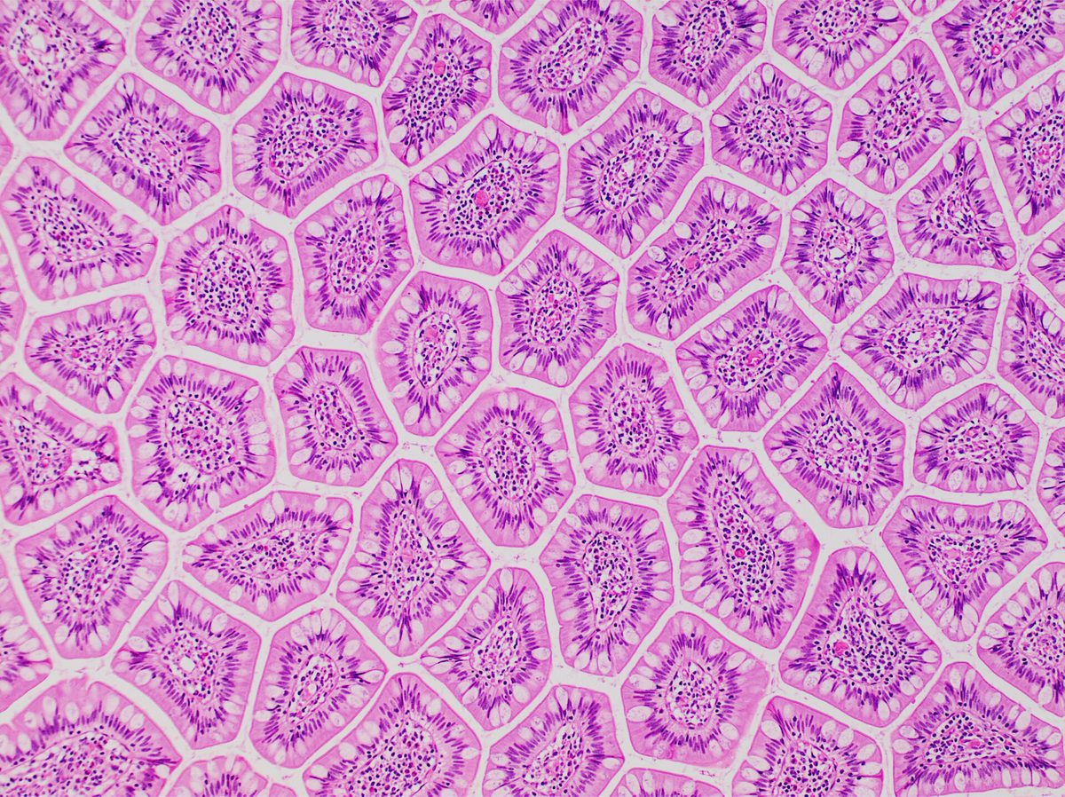
Absorption happens across a surface, so the rate depends on how much surface there is. A smooth tube a few metres long would offer a few hundred square centimetres. The small intestine instead has three levels of folding: large circular folds in the wall, finger-like villi standing up from those folds, and on each cell of each villus a dense brush of smaller projections. Each level multiplies the area, and together they reach something like thirty square metres.
This is the same geometrical idea that will reappear in the lungs and in the gills of a fish: when an exchange has to happen fast, evolution folds the surface. It is also the reason mechanical digestion matters on the food side — breaking one lump into many pieces increases the exposed surface without changing the mass at all.
Each villus also contains a capillary network and a lymph vessel, so an absorbed molecule has a very short distance to travel before it is carried away. Short distance and large area together are what make the rate possible; either on its own would not be enough.
Pause and explain
Why does the same mass of food digest faster when cut into small pieces?
Check your reasoning after answering
Because enzymes act at surfaces, and cutting a lump into pieces increases the exposed surface without changing the mass. More surface means more enzyme molecules can be in contact with the food at once, so the rate of breakdown rises. It is the same reason the intestinal lining is folded into villi — surface area governs the rate of a surface process.
Part 4 · From absorbed molecules to cellular work
Absorption is not the end of the story, and treating it as one is the error this unit most wants to prevent. Once glucose crosses into the blood it is transported, and it is only useful when it reaches a cell, crosses that cell's membrane, and is broken down in a mitochondrion with oxygen to release energy. Digestion supplies the raw material; the energy release happens somewhere else entirely, in the process from Chapter 2.
Amino acids follow a different route with the same shape. Absorbed into the blood, they are delivered to cells and used to build the proteins that cell needs, following its own DNA instructions. The protein in a meal is not transferred into your muscles; it is dismantled into amino acids and those are reassembled into entirely different proteins.
So an experiment that shows food breaking down faster has shown exactly that and nothing more. It has not shown that absorption occurred, that transport happened, or that any energy was released. Each stage needs its own evidence, which is the discipline from Chapter 1 applied to a body instead of a windowsill.
Pause and explain
Does protein eaten in a meal become muscle protein directly?
Check your reasoning after answering
No. Digestion cuts dietary protein into amino acids, which are absorbed and transported to cells. Each cell then builds its own proteins from those amino acids, in the order its own DNA specifies. The amino acids are reused; the original protein is not transferred, which is why one food protein can contribute to thousands of different body proteins.
Apply the whole unit · Interpret a digestion model
Illustrative comparison, not a measured experiment: Sample A is left in one large piece; Sample B has the same mass but is divided into small pieces. Both are placed under the same stated enzyme conditions, and B breaks down faster.
Your task
Explain the mechanism through surface area, distinguish what was observed from what is inferred, and connect the result to absorption.
What your response should show
- Explain the relevant biological process accurately.
- Use the supplied evidence and show calculation steps with units where needed.
- Connect a labeled model or mechanism with the claim.
- State an assumption, evidence limitation, or useful next observation.
Response guidance — after your attempt
State the mechanism: enzymes act where they touch the food, so the rate depends on exposed surface, and dividing a sample increases surface without changing mass.
Separate observation from inference: the observation is that B broke down faster under the stated conditions; the inference is that the greater exposed surface caused it.
Name what the result does not show: nothing about absorption, transport or energy release, which are separate stages needing separate evidence.
Connect to the body: this is why chewing precedes chemical digestion, and why the intestinal lining is folded into villi — the same surface-area principle on the food side and the gut side.
B breaking down faster is consistent with a surface-area mechanism. Enzymes work where they are in contact with their substrate, so the rate of chemical digestion depends on how much of the food is exposed. Dividing a sample into small pieces leaves the mass unchanged and multiplies the exposed surface, so more enzyme molecules can act at once and breakdown proceeds faster. The observation here is narrow and worth stating precisely: under the stated enzyme conditions, the divided sample broke down more quickly than the intact one. The mechanism is an inference, and a plausible rival worth checking would be that the act of dividing also warmed the sample or changed how the enzyme solution circulated around it, either of which could contribute. The result says nothing at all about the later stages. It does not show that any product was absorbed, that anything was transported, or that energy was released, because each of those happens elsewhere and would need its own evidence — a measurement of what crossed a membrane, or of oxygen consumed. In the body the same principle appears twice: chewing increases the food's surface before the enzymes reach it, and the intestinal lining is folded into villi and then into a brush of finer projections, reaching an absorbing area of roughly thirty square metres so that absorption can keep pace.
Scientific references for this unit
Parent / teacher teaching notes
Ask for a because-statement and a labeled relationship, not just a memorized word. Work through one calculation step at a time, permit oral or diagram responses, and use flexible sessions for difficult reading. Compare the learner’s actual evidence with the supplied model; do not reward copying or agreement with a personal preference. No private family, genetic, or medical records are required.
The key idea
Digestion breaks food into smaller components that can be absorbed and used. Mechanical processes such as chewing change size; chemical digestion changes large molecules into smaller molecules through reactions. Enzymes help these reactions occur. Digestion does not simply convert food directly into blood or energy.
Worked examples
Example 1 · Predict the effect of removing a contribution
- Identify what bile does: it emulsifies fat, breaking large droplets into many small ones — mechanical digestion, not chemical.
- Identify what lipase needs: it acts at the surface of fat droplets, so the rate depends on the total droplet surface available.
- Combine the two: without emulsification the fat stays in large droplets with much less surface, so lipase works far more slowly even though it is present in normal amounts.
A mechanical contribution can limit a chemical process. Ask what each step supplies.
Example 2 · Judge what an experiment showed
- State what was observed: starch was converted to sugar in the presence of saliva within ten minutes.
- Identify the mechanism that supports: amylase in saliva cuts the starch chain into smaller sugars — chemical digestion.
- Check the conclusion against the stages: energy release requires the sugar to enter a cell and be respired in a mitochondrion with oxygen, none of which happened in the test tube.
Each stage of the chain needs its own evidence. A test tube cannot show what a cell does.
Avoid these mistakes
- Mistake: chewing is enough to let food be absorbed. Chewing is mechanical — the starch is still a long chain; amylase has to cut it before anything can cross a membrane.
- Mistake: bile digests fat. Bile emulsifies fat into small droplets, which is mechanical; lipase does the chemical digestion, and it works faster because of the surface bile created.
- Mistake: eating protein builds muscle protein directly. Protein is cut into amino acids, absorbed, and reassembled by each cell into entirely different proteins its own DNA specifies.
- Mistake: faster breakdown shows energy was released. Breakdown, absorption, transport and respiration are four stages; an experiment showing one of them shows nothing about the other three.
Check your understanding
Why is grinding alone not complete chemical digestion?
Discuss the answer
It changes piece size and surface area but does not necessarily break the relevant chemical bonds into absorbable components.
Why is the stomach-only model incomplete?
Discuss the answer
Processing, transport, digestive contributions, and much absorption occur through several organs, especially the small intestine.
Words to know
- digestion
- Breaking food down, mechanically into smaller pieces and chemically into smaller molecules; only the chemical stage makes molecules small enough to absorb.
- enzyme
- A protein that cuts a particular kind of molecule — amylase on starch, protease on protein, lipase on fat — working within a range of acidity and temperature.
- absorption
- The movement of small food molecules out of the gut and into the blood, mostly across the lining of the small intestine.
- small intestine
- The organ where most chemical digestion is completed and most absorption happens, with enzymes delivered into it by the pancreas and bile by the liver.
- surface area
- How much surface is exposed; it governs the rate of any surface process, which is why food is chewed and the gut lining is folded.
Extend your scientific explanation
Compare a second supplied example or the linked educational reference. Identify a connection, a model limit, and an additional observation that would test the explanation.
In the real world
Use a clear question, reliable observations, and a biological mechanism to evaluate a claim. State what remains uncertain before applying a classroom model to a different setting.
Exit check
Does absorbing sugar mean all its energy has already been used?
Check your explanation after answering
No. Absorption makes material available for transport; later cellular processes transfer its stored energy or use its matter.
Explain your reasoning without notes. If a key idea is missing, revisit the lesson and try again.
Circulatory & Respiratory Systems
Learning goals
- I can explain circulatory & respiratory systems.
- I can use evidence, a model, and stated assumptions to support a biological explanation.
Before this unit
Use the supplied vocabulary, distinguish an observation from an explanation, and identify any units or model assumptions. Revisit a connected earlier unit if needed; new terms are explained here.
Use three flexible 40–45-minute sessions: core background and Parts 1–2 with their checks; Parts 3–4 and the worked application; then remaining practice, a labeled model, revision, and the exit check. Checks completed during reading can count toward the practice set. Read one short part at a time. Investigations and chapter review/quiz have separate sessions, and flexible weeks allow more time.
Hold your breath and the discomfort you feel within a minute is not a shortage of oxygen. It is a rise in carbon dioxide, which your body detects far more sensitively. That distinction is the right way into this unit, because it separates two things the word breathing runs together. Ventilation is the mechanical movement of air in and out of your lungs — ribs lifting, the diaphragm flattening, pressure dropping so that air flows in. Gas exchange is the chemical traffic that ventilation serves: oxygen crossing from air into blood, carbon dioxide crossing the other way. Ventilation is a pump; gas exchange is a transfer across a surface; and cellular respiration, from Chapter 2, is a third thing again, happening in mitochondria a long way from either.
Gas exchange happens across a surface built for it. Your lungs divide into roughly three hundred million tiny air sacs, each wrapped in blood vessels, and the resulting surface is somewhere around seventy square metres — comparable to a badminton court folded into your chest. The barrier between air and blood is about a thousandth of a millimetre thick. Large area, short distance, and a constant difference in concentration maintained at both ends: those are the same three conditions that made the small intestine's lining work, and they are what any fast exchange surface needs.
Delivering the gas is the circulatory system's job. Blood leaves the heart in an artery, which has thick muscular walls to withstand the pressure of each beat. It returns in a vein, which is wider, thinner-walled and carries valves to stop backflow in the low-pressure return. Between them lie the capillaries, so narrow that red blood cells pass almost single file, with walls one cell thick — and that is where every exchange with tissue actually happens. Arteries and veins are plumbing; capillaries are the point of the whole system. Blood runs in two connected circuits: heart to lungs and back, then heart to body and back, which is why the heart has four chambers rather than two.
Build your understanding
Where this fits
Digestion delivered food molecules to the gut wall and absorption put them into the blood. Energy release also needs oxygen, and oxygen enters somewhere else entirely. This unit covers the two systems that collect oxygen and deliver both it and the absorbed food to every cell — and separates breathing, which you can feel, from the chemistry it serves.
Use three flexible 40–45-minute sessions: core background and Parts 1–2 with their checks; Parts 3–4 and the worked application; then remaining practice, a labeled model, revision, and the exit check. Checks completed during reading can count toward the practice set. Read one short part at a time. Investigations and chapter review/quiz have separate sessions, and flexible weeks allow more time.
Part 1 · Breathing is not gas exchange, and neither is respiration
Three different things wear overlapping names, and separating them fixes most of the confusion in this topic. Ventilation moves air: the diaphragm contracts and flattens, the rib cage lifts, the volume of the chest increases, pressure inside falls below atmospheric, and air flows in. Nothing chemical has happened yet.
Gas exchange is the transfer at the air sac surface. Oxygen is more concentrated in the fresh air than in the blood arriving, so it diffuses into the blood; carbon dioxide is more concentrated in that blood than in the air, so it diffuses out. This is the diffusion from Chapter 2 — no pumping, no energy spent, just movement down concentration gradients across a thin wet surface.
Cellular respiration is the chemistry that creates those gradients in the first place. Cells consume oxygen and produce carbon dioxide, which keeps blood oxygen low and blood carbon dioxide high, which keeps the diffusion running at the lungs. Ventilation exists to keep the air side of the gradient fresh. A plant, which has no lungs and does not ventilate, still respires.
Pause and explain
Distinguish ventilation, gas exchange and cellular respiration in one sentence each.
Check your reasoning after answering
Ventilation is the mechanical movement of air into and out of the lungs, driven by the diaphragm and ribs changing the volume of the chest. Gas exchange is the diffusion of oxygen into the blood and carbon dioxide out of it, across the air sac surface. Cellular respiration is the chemical release of energy from glucose inside mitochondria, which is what consumes the oxygen and produces the carbon dioxide.
Part 2 · What an exchange surface has to be
Three features make a surface good at exchange, and the lungs have all three to an extreme degree. A large area: three hundred million air sacs reaching perhaps seventy square metres. A short diffusion distance: the wall between air and blood is roughly one micrometre, two cells thick. And a maintained concentration difference: ventilation refreshes the air on one side while circulation carries away the blood on the other, so the gradient never has a chance to even out.
That third condition is the one usually forgotten, and without it the other two would not help for long. If blood sat still in the capillaries it would reach equilibrium with the air and exchange would stop, exactly as the diffusion unit predicted. A continuous flow on both sides keeps the system permanently far from equilibrium, which is what keeps it moving.
The same three features appear wherever fast exchange is needed, which is a good sign that the explanation is right rather than a story about lungs. Fish gills have filaments and lamellae giving a huge area, a thin barrier, and water flowing one way past blood flowing the other. The small intestine has villi, a one-cell lining and a capillary carrying absorbed molecules away. Different organs, same requirements.
Pause and explain
Why would gas exchange stop if blood stopped flowing through the lung capillaries, even with the lungs still ventilating?
Check your reasoning after answering
Because the blood in the capillaries would quickly reach equilibrium with the air — taking up oxygen until its concentration matched, and giving up carbon dioxide until the same. With no difference left across the surface, diffusion would have no net direction. Flow on the blood side is what keeps the gradient steep, just as ventilation does on the air side.
Part 3 · Two circuits, and why the heart has four chambers
Blood makes two journeys, not one. In the first, the right side of the heart pumps blood low in oxygen to the lungs, where it takes up oxygen and releases carbon dioxide, and it returns to the left side. In the second, the left side pumps that oxygen-rich blood to the whole body, where it delivers oxygen and collects carbon dioxide, and it returns to the right side. Two circuits, each beginning and ending at the heart.
This is why four chambers are needed: two to receive returning blood and two to pump it out, with the two circuits kept separate so that oxygen-rich and oxygen-poor blood do not mix. The left side pumps against far more resistance, since it must reach the feet and the brain, and its muscular wall is correspondingly thicker — a structural difference you can see, explained by a functional demand.
Keeping the two sides straight also explains the vessels. An artery carries blood away from the heart and a vein carries it back, and that is the definition — not whether the blood is rich in oxygen. The pulmonary artery carries oxygen-poor blood to the lungs, and the pulmonary vein carries oxygen-rich blood back. Defining the vessels by direction rather than by content is what makes the exception stop being confusing.
Pause and explain
The pulmonary artery carries blood low in oxygen. Does that break the definition of an artery?
Check your reasoning after answering
No, because an artery is defined by direction: it carries blood away from the heart. The pulmonary artery carries oxygen-poor blood away from the right side towards the lungs, and the pulmonary vein brings oxygen-rich blood back. Defining arteries and veins by what they carry rather than which way they go is what creates the apparent exception.
Part 4 · Connecting transport to what cells actually do
Everything above is delivery. The purpose only appears at the capillary, where oxygen and glucose leave the blood, cross into tissue fluid, and pass through a cell's membrane; and where carbon dioxide makes the reverse trip. Capillary walls are one cell thick and the vessels are so fine that no cell in your body is far from one — that short distance is what makes diffusion fast enough to serve a working cell.
Inside the cell, the mitochondria consume the oxygen and the glucose and release the energy. This is the step people skip when they say that breathing gives you energy. Breathing moves air. The energy comes from a chemical reaction in cells, which the air movement supplies one reactant for, and which would also need the glucose that arrived by an entirely separate route through the digestive system.
A clean way to check your own explanation is to ask where each substance was at each stage and what moved it. Oxygen: air, then lung capillary blood, then heart, then body capillary, then tissue fluid, then cell, then mitochondrion. Each step has a mechanism — bulk flow driven by the heart, or diffusion down a gradient. If a step in your account has no mechanism, that is the part to work on.
Pause and explain
A draft says oxygen moves directly from the lungs to every muscle cell. What is missing?
Check your reasoning after answering
The whole transport stage. Oxygen diffuses from the air sacs into blood in the lung capillaries, is carried to the left side of the heart, pumped out through arteries, and reaches tissues through capillaries, where it diffuses into tissue fluid and then into cells. Only then is it used, in mitochondria. Without the heart and vessels there is no route.
Apply the whole unit · Repair a systems diagram
Illustrative draft, not a measured record: a student's diagram shows air entering the lungs, then an arrow labelled oxygen going straight from the lungs to every muscle cell. No vessels and no heart are drawn.
Your task
Repair the pathway, label where each gas exchange happens, and distinguish the pathway from cellular respiration.
What your response should show
- Explain the relevant biological process accurately.
- Use the supplied evidence and show calculation steps with units where needed.
- Connect a labeled model or mechanism with the claim.
- State an assumption, evidence limitation, or useful next observation.
Response guidance — after your attempt
Insert the missing stage: oxygen diffuses from the air sacs into blood in the lung capillaries, which the draft omits entirely.
Add the transport: that blood returns to the left side of the heart and is pumped through arteries to capillaries throughout the body.
Label the second exchange: at a body capillary, oxygen diffuses into tissue fluid and then into cells, while carbon dioxide diffuses the other way and is carried back for release at the lungs.
Separate the chemistry: cellular respiration happens inside the cells' mitochondria, consuming the delivered oxygen and glucose — it is not part of the transport pathway.
The draft is missing everything between the lungs and the cells. Repaired, the pathway runs: air is drawn into the air sacs by ventilation; oxygen diffuses across the thin sac wall into blood in the surrounding capillaries, because oxygen is more concentrated in fresh air than in the arriving blood; that blood returns to the left side of the heart and is pumped out through arteries, which branch until they become capillaries in the tissues; at a body capillary oxygen diffuses out into tissue fluid and across the membranes of nearby cells. Carbon dioxide follows the reverse route — produced in cells, diffusing into the capillary blood, carried back to the right side of the heart, pumped to the lungs, and diffusing into the air sacs to be removed by ventilation. Two exchanges, both by diffusion down concentration gradients, with bulk flow driven by the heart in between. Cellular respiration is a separate thing and belongs inside the cell: it is the reaction in which mitochondria use the delivered oxygen and glucose to release energy, and it is what produces the carbon dioxide and keeps both gradients steep. The draft's single arrow collapses a pump, two exchange surfaces and a chemical reaction into one step. Worth noting too that the glucose arrives by a different route entirely, absorbed in the small intestine and carried by the same circulation.
Scientific references for this unit
Parent / teacher teaching notes
Ask for a because-statement and a labeled relationship, not just a memorized word. Work through one calculation step at a time, permit oral or diagram responses, and use flexible sessions for difficult reading. Compare the learner’s actual evidence with the supplied model; do not reward copying or agreement with a personal preference. No private family, genetic, or medical records are required.
The key idea
The respiratory system moves air and exchanges gases; the circulatory system transports materials between organs and cells. In lungs, oxygen moves from air spaces into blood while carbon dioxide can move in the other direction. The heart pumps blood through vessels. These tasks interact, but ventilation, gas exchange, and cellular respiration are distinct processes.
Worked examples
Example 1 · Explain a structure from a demand
- Identify what each side pumps to: the right side sends blood to the lungs, which are close by; the left side sends it to the entire body, including the brain and feet.
- Compare the resistance: a long branching network reaching the extremities offers far more resistance than the short pulmonary circuit.
- Connect structure to demand: generating the higher pressure needed requires more muscle, so the left wall is thicker.
A visible structural difference usually has a functional demand behind it. Name the demand.
Example 2 · Find the missing mechanism
- List the stages actually required: ventilation, gas exchange at the lungs, transport by blood, exchange at a body capillary, and cellular respiration.
- Attach a mechanism to each: pressure difference for ventilation, diffusion for both exchanges, bulk flow driven by the heart for transport, and enzyme-controlled chemistry in mitochondria.
- Note the second reactant: glucose from digestion, delivered by the same circulation, without which oxygen releases nothing.
If a step in an account has no mechanism, that is the part that needs work.
Avoid these mistakes
- Mistake: breathing is respiration. Ventilation moves air mechanically; cellular respiration is a chemical reaction in mitochondria. A plant respires and does not breathe.
- Mistake: arteries carry oxygen-rich blood by definition. They carry blood away from the heart — the pulmonary artery carries oxygen-poor blood to the lungs.
- Mistake: oxygen goes straight from lungs to muscles. It crosses into blood, travels through the heart and arteries, and diffuses out at a capillary; without the transport stage there is no route.
- Mistake: a big surface is enough for fast exchange. Area, a short diffusion distance and a maintained concentration difference are all needed — blood that stopped flowing would reach equilibrium and exchange would halt.
Check your understanding
Is exhaled air pure carbon dioxide?
Discuss the answer
No. It remains a mixture including nitrogen, oxygen, carbon dioxide, and water vapor in different proportions.
Why are thin surfaces relevant?
Discuss the answer
They shorten the distance materials must cross and support exchange under suitable conditions.
Words to know
- ventilation
- The mechanical movement of air in and out of the lungs as the diaphragm and ribs change the volume of the chest; nothing chemical happens yet.
- gas exchange
- Diffusion of oxygen into the blood and carbon dioxide out of it across a thin, moist surface with a maintained concentration difference.
- capillary
- A vessel one cell thick and barely wider than a red blood cell, where every exchange with tissue actually happens.
- artery
- A vessel carrying blood away from the heart, with thick muscular walls for the pressure of each beat — defined by direction, not by oxygen content.
- vein
- A vessel carrying blood back towards the heart, wider and thinner-walled, with valves preventing backflow at low pressure.
Extend your scientific explanation
Compare a second supplied example or the linked educational reference. Identify a connection, a model limit, and an additional observation that would test the explanation.
In the real world
Use a clear question, reliable observations, and a biological mechanism to evaluate a claim. State what remains uncertain before applying a classroom model to a different setting.
Exit check
Why is inhalation alone not the whole oxygen pathway?
Check your explanation after answering
Oxygen must cross exchange surfaces, travel in blood, and reach cells; inhalation is only one step.
Explain your reasoning without notes. If a key idea is missing, revisit the lesson and try again.
Bones, Muscles & Structure-Function Models
Learning goals
- I can explain bones, muscles & structure-function models.
- I can use evidence, a model, and stated assumptions to support a biological explanation.
Before this unit
Use the supplied vocabulary, distinguish an observation from an explanation, and identify any units or model assumptions. Revisit a connected earlier unit if needed; new terms are explained here.
Use three flexible 40–45-minute sessions: core background and Parts 1–2 with their checks; Parts 3–4 and the worked application; then remaining practice, a labeled model, revision, and the exit check. Checks completed during reading can count toward the practice set. Read one short part at a time. Investigations and chapter review/quiz have separate sessions, and flexible weeks allow more time.
Pick up a pencil and watch the back of your hand. The muscles that move your fingers are not in your fingers — they are mostly in your forearm, pulling on cords that run across the wrist. That is why your fingers can be slim enough to be useful. It is a design decision visible in your own hand, and it introduces the three parts of this unit: bones that provide rigid levers, skeletal muscle that can only pull, and tendons, the tough cords that transmit that pull from a muscle to a bone, sometimes across a considerable distance.
Muscle has one trick. Contraction shortens it and generates a pull; relaxing lets it be stretched again but produces no push. A single muscle can therefore move a bone one way and has no way of returning it, which is why skeletal muscles work in opposing pairs. Your biceps contracts to bend the elbow and your triceps contracts to straighten it, and each one stretches the other as it works. Every movement of a joint — the place where bones meet and motion is possible — is produced by one of a pair shortening while its partner lengthens.
Put a rigid bone, a pivot at a joint, and a pull from a muscle together and you have a lever. In your forearm the elbow is the pivot, the forearm bone is the lever, the biceps supplies the effort close to the pivot, and the load sits far out at your hand. That arrangement is poor at force and excellent at speed and range: a small shortening of the biceps swings your hand through a large arc quickly, at the cost of having to pull many times harder than the weight you are lifting. Most of your limbs are built this way, trading force for speed, which tells you something about what bodies are usually for.
Build your understanding
Where this fits
Materials now reach the cells that need them. This unit asks what those cells do with the energy in one particular case: movement. It is also the chapter's clearest example of structure explaining function, and of how far a physical model of the body can be trusted before it starts to mislead.
Use three flexible 40–45-minute sessions: core background and Parts 1–2 with their checks; Parts 3–4 and the worked application; then remaining practice, a labeled model, revision, and the exit check. Checks completed during reading can count toward the practice set. Read one short part at a time. Investigations and chapter review/quiz have separate sessions, and flexible weeks allow more time.
Part 1 · Three structures, three jobs
Bone is rigid, which is what makes leverage possible, and it is not inert — it is living tissue with cells, a blood supply and the ability to repair and remodel. Its rigidity comes from mineral deposits, and its resistance to shattering from protein fibres woven through them, a composite rather like reinforced concrete.
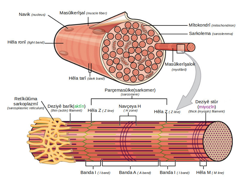
Skeletal muscle is built of long fibres that shorten when stimulated, and it is attached at both ends. One attachment is usually to a bone that stays relatively fixed and the other to the bone that moves. Tendons make those attachments: dense cords of collagen, barely stretchy, so that a muscle's shortening is transmitted rather than absorbed. A stretchy tendon would waste the contraction.
Joints are where motion becomes possible, and their shape sets the motion allowed. A hinge at the elbow permits bending in one plane; a ball and socket at the shoulder permits rotation in many; the bones of the skull are joined so firmly that no movement occurs at all. Cartilage at the surfaces and fluid in the cavity reduce friction and absorb shock. Structure, in each case, is a statement about what is permitted.
Pause and explain
Why would a stretchy tendon be a problem?
Check your reasoning after answering
Because a tendon's job is to transmit a muscle's shortening to a bone. If the tendon stretched, part of the contraction would be used extending the tendon rather than moving the bone, so the movement would be smaller, later and less precise. Tendons are made of dense, barely stretchy collagen for exactly that reason.
Part 2 · Muscles pull, which is why they come in pairs
A contracting muscle shortens and pulls. It cannot lengthen itself, and it cannot push. Everything about how limbs are arranged follows from that single limitation. To move a bone back the way it came, a second muscle is needed on the opposite side, and its contraction both produces the return movement and restores the first muscle to its original length.
The biceps and triceps are the standard pair. The biceps crosses the front of the elbow, so contracting it bends the joint; the triceps crosses the back, so contracting it straightens the joint. When one contracts the other relaxes and is stretched — and when you hold a position steady, both contract at once with their pulls balanced, which is why holding a weight still is tiring even though nothing moves.
Coordination comes from outside the muscles. Nerves carry the signals that time the contractions, and the next chapter takes that up. Here the point is structural: opposing pairs are not a redundancy but a necessary consequence of a tissue that can only pull, and the same arrangement appears everywhere, from your jaw to a bird's wing.
Pause and explain
Why can't the biceps straighten the arm by relaxing?
Check your reasoning after answering
Because relaxing only allows the muscle to be stretched; it generates no force. Something has to do the stretching, and that is the triceps on the opposite side of the elbow, which contracts to straighten the joint and lengthens the biceps as it does. A muscle that can only pull requires a partner that pulls the other way.
Part 3 · Using a lever model, and stating its assumptions
The forearm is usefully modelled as a rigid bar on a pivot. Three things matter: where the pivot is, where the effort is applied, and where the load acts. At the elbow the biceps attaches only a few centimetres from the joint while a weight in the hand is perhaps thirty centimetres away, so the muscle works at a large mechanical disadvantage and must pull several times harder than the weight it lifts.
What the body buys with that disadvantage is speed and range. A small shortening of the biceps near the pivot swings the hand through a wide arc, and quickly. For an animal that needs to throw, run or catch, that trade is worth making, and limbs built the other way — with the muscle attached far from the joint — are found where force matters more than speed, as in a digging mole's forelimb.
The model earns its keep and then has to be bounded. It treats the bone as weightless and perfectly rigid, the pivot as frictionless, and the muscle's pull as a single force of constant size acting at one point. None is true. Real muscle force varies with how stretched it is and how fast it is shortening, the attachment covers an area, and the joint has friction and soft tissue. The model predicts the direction of the trade well and the exact numbers poorly.
Pause and explain
The biceps attaches close to the elbow. What does the body gain and lose from that arrangement?
Check your reasoning after answering
It loses force efficiency: because the muscle acts close to the pivot while the load is far from it, the biceps must pull several times harder than the weight being lifted. It gains speed and range — a small contraction near the pivot moves the hand through a large arc quickly, which is worth more to an animal that throws, runs or catches than raw lifting force would be.
Part 4 · Movement depends on every other system
A contraction costs energy, and the energy comes from respiration in the muscle cells' mitochondria, using glucose delivered by the circulation from the digestive system and oxygen delivered from the lungs. Working muscle also produces heat and carbon dioxide faster than resting muscle, which is why exercise makes you warm and makes you breathe harder.
The signal to contract comes from the nervous system, and its timing is what turns a set of pulls into a movement rather than a spasm. Bone itself depends on the blood supply for the materials to repair and remodel, and on the hormonal system for the regulation of the minerals it is built from.
So the paper-and-string models used to demonstrate a joint show one thing honestly — that two opposing pulls can rotate a hinge in two directions — and leave out everything that makes the real system work: living tissue, energy supply, nerves, variable force and the changing geometry as a joint moves through its range. A model is worth using and worth bounding, which is the habit from Chapter 1 applied to an arm.
Pause and explain
Name two systems a single arm movement depends on, and what each supplies.
Check your reasoning after answering
The circulatory system, which delivers the glucose absorbed by the digestive system and the oxygen taken up by the lungs, so the muscle's mitochondria can release the energy contraction costs. And the nervous system, which carries the signals that time the contractions of the opposing pair, turning separate pulls into a controlled movement.
Apply the whole unit · Analyze an opposing-pull model
Illustrative paper model, not a measured apparatus: a card hinge has two strings attached. Pulling string A bends the hinge; pulling string B straightens it. The hinge and the attachment points are unchanged throughout.
Your task
Explain the mechanism the model represents, connect it to muscles at a real joint, and state two limits of the analogy.
What your response should show
- Explain the relevant biological process accurately.
- Use the supplied evidence and show calculation steps with units where needed.
- Connect a labeled model or mechanism with the claim.
- State an assumption, evidence limitation, or useful next observation.
Response guidance — after your attempt
Identify what the strings represent: two pulls applied on opposite sides of a pivot, each able to rotate the hinge one way.
Map it onto the body: string A corresponds to a muscle such as the biceps crossing the front of the elbow, string B to the triceps crossing the back.
State why two are needed: a muscle, like a string, can only pull, so a second one on the opposite side is required to reverse the movement and restore the first to length.
Name limits: the model has no energy supply, no nerves, no living tissue, a constant pull rather than a force that varies with length and speed, and no realistic anatomical scale.
The model represents a pair of opposing pulls acting on a pivot. Each string can only pull, and because they attach on opposite sides of the hinge, pulling A rotates it one way and pulling B rotates it back — which is exactly the arrangement of skeletal muscles at a joint. At the elbow the biceps crosses the front and bends the joint when it contracts; the triceps crosses the back and straightens it. Each one stretches the other as it works, which is the only way a tissue that can only shorten can produce movement in two directions. The model is honest about that mechanism and silent about everything else. It has no energy supply, where a real contraction is paid for by respiration in the muscle cells using glucose and oxygen delivered by two other systems. It has no nervous control, where real timing and the graded strength of a contraction come from nerve signals. Beyond those, the strings pull with whatever force you apply, while real muscle produces a force that varies with how stretched it is and how fast it is shortening; the attachments are points, while real tendons attach over an area; and the paper is not living tissue that repairs, fatigues and remodels. The model is adequate for explaining why muscles come in pairs and inadequate for predicting how hard a biceps pulls, which is the right way to judge any model: against the question being asked.
Scientific references for this unit
Parent / teacher teaching notes
Ask for a because-statement and a labeled relationship, not just a memorized word. Work through one calculation step at a time, permit oral or diagram responses, and use flexible sessions for difficult reading. Compare the learner’s actual evidence with the supplied model; do not reward copying or agreement with a personal preference. No private family, genetic, or medical records are required.
The key idea
Animal movement involves support structures, muscles, joints, and coordination. Bones provide support, protect some organs, and form parts of movable arrangements. Skeletal muscles contract and pull through connections such as tendons. A contraction can produce movement only in relation to the structures and forces involved.
Worked examples
Example 1 · Explain an arrangement from its trade-off
- Recall the lever trade: effort applied far from the pivot gives more force and less speed; applied close to the pivot it gives more speed and range and less force.
- Match each animal to what it needs: a mole digs through soil, which demands force against resistance rather than quick wide movements.
- Match the other: a cheetah needs its limbs to swing through a long arc very fast, and can accept that its muscles must pull much harder than the load.
The same lever principle explains both; only the demand differs.
Example 2 · Bound a model before trusting a number
- Check the direction of the result: the muscle acts far closer to the pivot than the load, so a force several times the weight is the right shape of answer.
- List what the model assumed: a weightless rigid bone, a frictionless pivot, a single point of attachment, and a pull of constant size.
- Decide what to claim: the calculation establishes the order of magnitude and the direction of the trade, not an accurate physiological value.
State what a model is adequate for. Here: the direction of the trade, not the precise number.
Avoid these mistakes
- Mistake: a muscle pushes to straighten a joint. Muscles only pull; straightening the elbow is the triceps contracting, not the biceps pushing.
- Mistake: relaxing a muscle moves the bone back. Relaxing generates no force — the opposing muscle of the pair does the work and stretches the relaxed one.
- Mistake: a limb lever is built for lifting power. Most limbs attach the muscle close to the joint, trading force for speed and range, which is why the biceps pulls far harder than the weight it lifts.
- Mistake: a paper hinge model shows how an arm works. It shows why opposing pairs are needed and omits energy supply, nerves, living tissue and the way real muscle force varies with length and speed.
Check your understanding
Why is listing bones alone not a structure-function explanation?
Discuss the answer
It identifies parts but does not explain how their arrangement contributes to support, protection, or movement.
Why are opposing muscle actions useful?
Discuss the answer
They allow movement in different directions because a contracting muscle pulls rather than actively pushes along the same attachment.
Words to know
- skeletal muscle
- Muscle attached to bones that shortens when stimulated, generating a pull; it cannot push, which is why muscles work in opposing pairs.
- tendon
- A dense, barely stretchy cord of collagen attaching muscle to bone, so that a contraction is transmitted rather than absorbed.
- joint
- A place where bones meet and movement is possible; its shape sets what movement is allowed — a hinge at the elbow, a ball and socket at the shoulder.
- contraction
- The shortening of a muscle that generates force; relaxing produces no push, only the possibility of being stretched by a partner.
- lever
- A rigid bar turning about a pivot; limbs trade force for speed and range by attaching the muscle close to the joint.
Extend your scientific explanation
Compare a second supplied example or the linked educational reference. Identify a connection, a model limit, and an additional observation that would test the explanation.
In the real world
Use a clear question, reliable observations, and a biological mechanism to evaluate a claim. State what remains uncertain before applying a classroom model to a different setting.
Exit check
Why does movement depend on more than muscles?
Check your explanation after answering
Coordination, support, exchange, and cellular energy processes interact with muscle contraction.
Explain your reasoning without notes. If a key idea is missing, revisit the lesson and try again.
Kidneys, Excretion & Internal Balance
Learning goals
- I can explain kidneys, excretion & internal balance.
- I can use evidence, a model, and stated assumptions to support a biological explanation.
Before this unit
Use the supplied vocabulary, distinguish an observation from an explanation, and identify any units or model assumptions. Revisit a connected earlier unit if needed; new terms are explained here.
Use three flexible 40–45-minute sessions: core background and Parts 1–2 with their checks; Parts 3–4 and the worked application; then remaining practice, a labeled model, revision, and the exit check. Checks completed during reading can count toward the practice set. Read one short part at a time. Investigations and chapter review/quiz have separate sessions, and flexible weeks allow more time.
Your kidneys filter something like 180 litres of fluid a day, and you produce roughly 1.5 litres of urine. Almost everything that goes through the filter is taken straight back. That ratio is the whole design, and it looks wasteful until you see what it buys: rather than trying to pick individual waste molecules out of the blood, the kidney pushes nearly everything small out of the blood and then deliberately reclaims what it wants to keep. Sorting by removal and selective return turns out to be far easier than sorting by selection, and it is why the kidney can handle substances it has never encountered before.
The vocabulary separates two things people run together. Excretion is the removal of waste products made by the body's own chemistry — carbon dioxide from respiration, urea from breaking down surplus amino acids. Egestion, passing undigested food out of the gut, is not excretion at all, because that material never took part in the body's metabolism; it passed through. Several organs excrete: lungs remove carbon dioxide, skin loses some water and salts in sweat, and the kidneys remove urea and adjust water and salt.
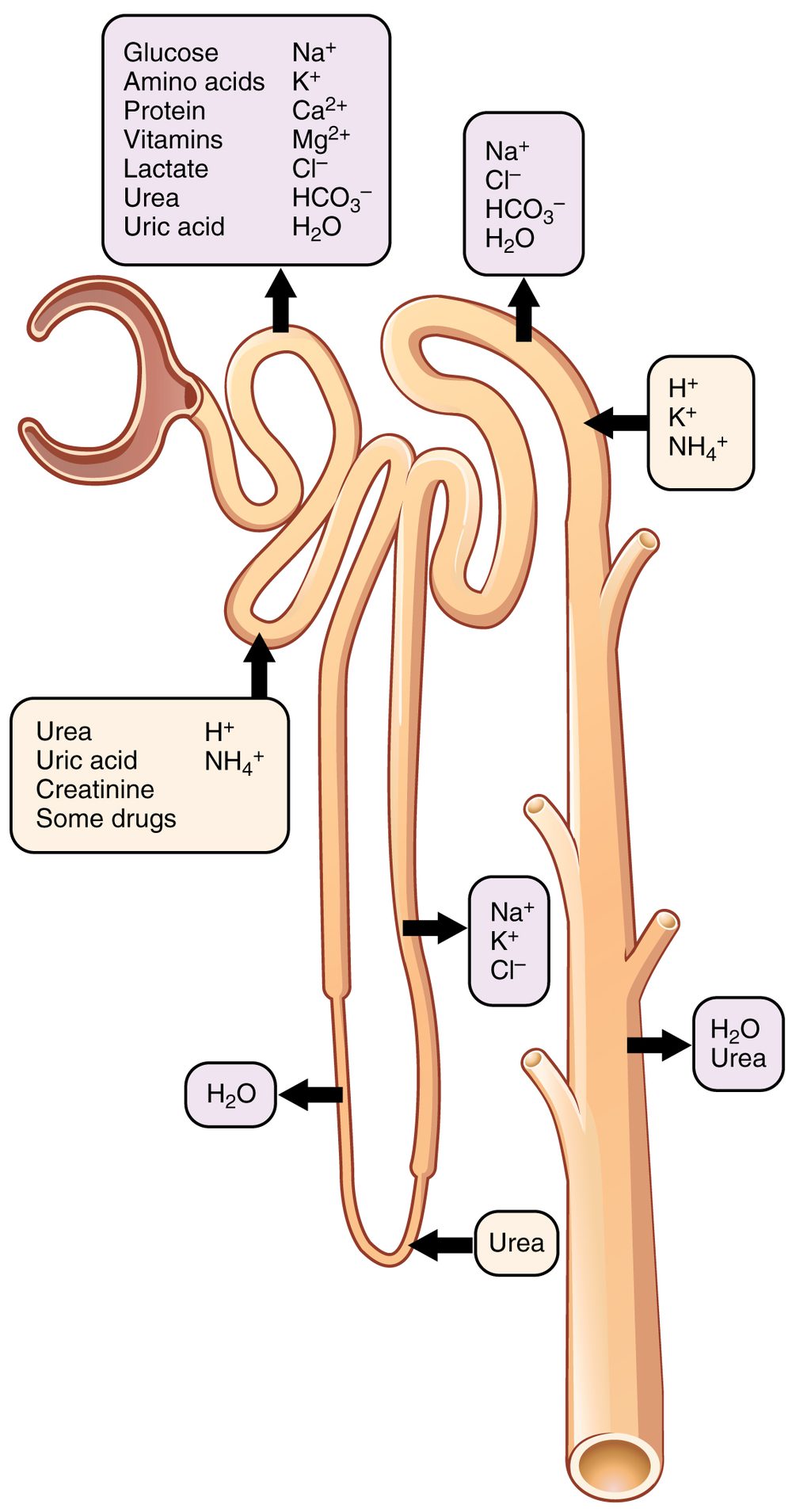
The kidney does its work in two stages. Filtration forces water and small dissolved molecules out of the blood under pressure, leaving blood cells and large proteins behind because they are too big to pass. What crosses is called filtrate, and it contains useful things and waste together — glucose, salts, water and urea all go through. Then comes reabsorption: as the filtrate travels along a tubule, the useful substances are taken back into the blood, glucose almost entirely, water and salts in amounts the body adjusts. What is left at the end is urine. The adjustment is the point — on a hot day with little to drink, far more water is reabsorbed and the urine is concentrated.
Build your understanding
Where this fits
This chapter has followed materials in: food broken down and absorbed, oxygen collected, both delivered, and energy released in cells doing work. Every one of those reactions produces something the body has to get rid of. This unit closes the chapter with the system that removes waste while keeping what is still useful — and it introduces the balance-sheet reasoning the ecology chapter will use again.
Use three flexible 40–45-minute sessions: core background and Parts 1–2 with their checks; Parts 3–4 and the worked application; then remaining practice, a labeled model, revision, and the exit check. Checks completed during reading can count toward the practice set. Read one short part at a time. Investigations and chapter review/quiz have separate sessions, and flexible weeks allow more time.
Part 1 · Which wastes leave by which route
Carbon dioxide is produced by respiration in every cell, diffuses into the blood, and is removed at the lungs. It is a genuine excretory product, and the lungs are therefore an excretory organ as well as a gas exchange surface — which surprises people who associate excretion only with the kidneys.
Urea comes from a different source. Surplus amino acids cannot be stored, so the liver breaks them down, and the nitrogen-containing part becomes urea, which is toxic if it accumulates. The liver makes it, the blood carries it, and the kidney removes it. Three organs in three systems for one waste product, which is a good illustration of why organs are grouped by function rather than by location.
Undigested food is the case that does not belong. Fibre and other material that passed through the gut without being absorbed never entered the body's chemistry, so passing it out is egestion, not excretion. The distinction is not a word game: it tells you where to look for a cause when something goes wrong, because an excretory problem and a digestive one have completely different origins.
Pause and explain
Why is passing undigested food out of the gut not excretion?
Check your reasoning after answering
Because excretion is the removal of waste produced by the body's own metabolism, and undigested food never took part in it — it passed through the gut without being absorbed into cells or reactions. Carbon dioxide from respiration and urea from breaking down surplus amino acids are excretory products; fibre is not.
Part 2 · Filter almost everything, then take back what you want
Filtration happens at a dense knot of capillaries under high pressure. The pressure drives water and small dissolved molecules out through a barrier that holds back blood cells and large proteins purely by size. Nothing is being selected at this stage: glucose, salts, amino acids, water and urea all cross together, which is why the volume filtered is so enormous.
Selection happens afterwards, along the tubule. Glucose is reabsorbed essentially completely, because losing it would waste the output of digestion. Salts and water are reabsorbed in amounts that vary with what the body needs. Urea is largely left behind, since removing it is the point of the exercise. By the end of the tubule the filtrate has been transformed from something resembling blood plasma into urine.
Reabsorption is not free. Taking glucose and salts back across into the blood often means moving them from a lower concentration to a higher one, which is the active transport from Chapter 2 — it needs pumps and it needs energy from respiration. The kidney is among the most energy-hungry organs in the body for exactly this reason, and that cost is what buys the flexibility of filtering first and sorting later.
Pause and explain
Why does the kidney filter far more fluid than it excretes?
Check your reasoning after answering
Because filtering is unselective: pressure pushes water and all small dissolved molecules out of the blood together, waste and useful substances alike. The sorting is done afterwards by reabsorbing what the body wants to keep. Filtering broadly and reclaiming selectively is simpler than picking individual waste molecules out, and it works on substances the body has never met before.
Part 3 · Tracking amounts within a stated boundary
Any system that takes something in and lets something out can be checked with a balance sheet, and doing so catches errors quickly. Draw a boundary — here, the processing of one batch of filtrate. Count what enters. Count what leaves by each route. If the two do not match, either something is accumulating inside the boundary or a route has been missed.
Work the model. Suppose 100 units of water enter the filtrate, 96 return to the blood, and 4 leave in the urine. Input is 100; output is 96 + 4 = 100; the model balances, and nothing is accumulating. The fraction returned is 96 ÷ 100 = 96 per cent, which says that almost all the filtered water is reclaimed — the central fact about how the kidney works.
The discipline matters more than the arithmetic. If the same model had shown 96 returning and 8 leaving, the total would be 104 against an input of 100, and you would know immediately that something was wrong with the figures rather than with the kidney. The same check will reappear with carbon and water in the ecology chapter, where it is the main defence against a plausible-sounding but impossible account.
Pause and explain
In the model, 100 units enter the filtrate, 96 return to the blood and 4 leave in the urine. Does the model balance, and what fraction returns?
Check your reasoning after answering
Yes. The output is 96 + 4 = 100 units, matching the 100 that entered, so nothing is unaccounted for and nothing is accumulating inside the boundary. The fraction returned is 96 ÷ 100, which is 96 per cent — almost all of the filtered water is reabsorbed, which is the central feature of how the kidney works.
Part 4 · Regulation, and what an invented model cannot tell you
How much water is reabsorbed is not fixed. Sensors detect how concentrated the blood is; a hormone released in response adjusts the permeability of part of the tubule; more water is reclaimed and the urine becomes concentrated. Drink a litre of water and the reverse happens within the hour. This is the negative feedback the next chapter takes up, with the kidney as its clearest case: a deviation is detected and a response reduces it.
Notice how many systems one piece of regulation involves. The circulatory system delivers the blood and carries the hormone; the hormonal system produces it; the nervous system contributes to the thirst that changes behaviour; and the kidney does the work. A regulated quantity is maintained by a loop running through several systems, not by one organ deciding something.
Finally, be clear about what a teaching model is. The 100-unit model above is invented to show the structure of filtration and selective return. Its numbers are chosen for arithmetic, not measured, and the real figures depend on body size, temperature, activity and what a person has drunk. It is not a statement about how much anyone should drink, and a model that balances neatly is evidence that the model is consistent, not that it is true of any particular body.
Pause and explain
On a hot day with little to drink, what changes in the kidney, and what detects the need?
Check your reasoning after answering
More water is reabsorbed along the tubule, so less leaves and the urine is more concentrated. The need is detected by sensors responding to how concentrated the blood has become; a hormone released in response increases the permeability of part of the tubule so more water returns to the blood. That is negative feedback: the deviation triggers the response that reduces it.
Apply the whole unit · Balance a supplied water model
Illustrative processing record for one model interval, not measured data: 100 water units enter the filtrate, 96 return to the blood pathway, and 4 leave through the urine pathway.
Your task
Check that the model balances, calculate the fraction returned, and explain why these figures are not a recommendation about how much anyone should drink.
What your response should show
- Explain the relevant biological process accurately.
- Use the supplied evidence and show calculation steps with units where needed.
- Connect a labeled model or mechanism with the claim.
- State an assumption, evidence limitation, or useful next observation.
Response guidance — after your attempt
Add the outputs: 96 units returned plus 4 units leaving gives 100 units, which matches the 100 that entered, so the model balances and nothing is unaccounted for.
Calculate the returned fraction: 96 ÷ 100 = 0.96, or 96 per cent of the filtered water reabsorbed.
Interpret the figure: it shows selective return rather than wholesale excretion, which is the central feature of kidney function.
State the limits: the numbers are invented for one stated boundary and one interval, and real values vary with body size, temperature, activity and intake.
The model balances. Its 100 input units are accounted for exactly by 96 units returning to the blood and 4 leaving in the urine, which sum to 100, so nothing is accumulating inside the boundary and no route has been missed. The returned fraction is 96 ÷ 100, which is 96 per cent. What that figure illustrates is the kidney's basic strategy: filtration is unselective, pushing water and all small dissolved molecules out of the blood together, and the sorting happens afterwards as reabsorption reclaims almost all of the water along with the glucose and the salts the body wants to keep. Only a small remainder, carrying the urea, leaves as urine. This is also why the kidney consumes so much energy, since much of that reabsorption is active transport working against a concentration gradient. The figures cannot be read as advice. They are invented for one stated boundary over one model interval, chosen so the arithmetic is clear, and a real kidney's filtered volume and reabsorbed fraction depend on body size, temperature, activity, health and how much a person has drunk. A model balancing neatly shows that the model is internally consistent, which is a different claim from being true of any particular body. What would move this towards a measurement is a stated interval, a stated body, and values obtained rather than assumed.
Scientific references for this unit
Parent / teacher teaching notes
Ask for a because-statement and a labeled relationship, not just a memorized word. Work through one calculation step at a time, permit oral or diagram responses, and use flexible sessions for difficult reading. Compare the learner’s actual evidence with the supplied model; do not reward copying or agreement with a personal preference. No private family, genetic, or medical records are required.
The key idea
Cells produce waste materials through metabolism, and organisms regulate internal water and dissolved substances. Excretion removes metabolic wastes; it differs from elimination of unabsorbed food from the digestive tract. Lungs release carbon dioxide, while kidneys help remove some dissolved wastes and regulate blood composition.
Worked examples
Example 1 · Find the missing route in a balance
- Add the stated outputs: 90 + 4 = 94 units leaving by the two named routes.
- Compare with the input: 100 units entered, so 6 units are unaccounted for.
- Interpret: either a route is missing from the model, or something is accumulating inside the boundary, or one of the figures is wrong.
Inputs must equal outputs plus accumulation. When they do not, suspect the model first.
Example 2 · Predict a change from a demand
- Identify the regulated quantity: the concentration of the blood, which rises when water is lost and falls when water is taken in.
- Apply the response for the dehydrated person: sensors detect more concentrated blood, a hormone increases water reabsorption along the tubule, so less water leaves.
- Apply the reverse for the other: dilute blood means less reabsorption, so more water leaves.
Name the detected quantity and the response that reduces the deviation — that is the shape of negative feedback.
Avoid these mistakes
- Mistake: passing faeces is excretion. Undigested food never entered the body's metabolism, so that is egestion; carbon dioxide and urea are excretory products.
- Mistake: only the kidneys excrete. The lungs remove carbon dioxide and the skin loses water and salts in sweat, so both are excretory organs too.
- Mistake: the kidney selects waste out of the blood. It filters almost everything small out indiscriminately and then reabsorbs what it wants to keep — in the model, 96 of every 100 water units.
- Mistake: a model that balances is a measurement. The 100-unit model is invented so the arithmetic is clear; real volumes depend on body size, temperature, activity and intake, and it is not advice about drinking.
Check your understanding
Why is excretion not identical to eliminating unabsorbed food?
Discuss the answer
Excretion removes wastes produced by metabolism; unabsorbed food is material that did not enter those cellular pathways.
Does entering a filtrate guarantee a substance leaves in urine?
Discuss the answer
No. Some substances are returned to blood during subsequent processing.
Words to know
- excretion
- Removing waste made by the body's own metabolism — carbon dioxide from respiration, urea from surplus amino acids — which is not the same as passing undigested food out.
- filtration
- Forcing water and small dissolved molecules out of the blood under pressure, holding back cells and large proteins by size alone; nothing is selected at this stage.
- reabsorption
- Taking useful substances back from the filtrate into the blood, often by active transport against a gradient, which is why the kidney is so energy-hungry.
- urine
- What remains of the filtrate after reabsorption: mostly water with urea and surplus salts, concentrated or dilute according to what the body needs.
- metabolism
- The chemical reactions going on inside an organism; excretion removes their products, which is why material that merely passed through the gut does not count.
Extend your scientific explanation
Compare a second supplied example or the linked educational reference. Identify a connection, a model limit, and an additional observation that would test the explanation.
In the real world
Use a clear question, reliable observations, and a biological mechanism to evaluate a claim. State what remains uncertain before applying a classroom model to a different setting.
Exit check
Does regulation require an unchanging number at every instant?
Check your explanation after answering
No. Regulated conditions can vary within ranges while feedback and exchanges help maintain function.
Explain your reasoning without notes. If a key idea is missing, revisit the lesson and try again.
Problem 1
Answer and explanation
The digestive system processes and absorbs food molecules, the circulatory system transports them, and muscle cells use them in cellular work. Organs contain interacting tissues, and tissues contain specialized cells. The pathway connects levels and systems; neither an intestine nor a muscle cell independently performs every step. The diagram is a selected model, not a complete anatomical description.
Identify digestive and circulatory organ-system roles.
Locate exchange surfaces within organs and tissues.
Connect transport to cellular use.
Response criteria
- Explain the relevant biological process accurately.
- Use the supplied evidence and show calculation steps with units where needed.
- Connect a labeled model or mechanism with the claim.
- State an assumption, evidence limitation, or useful next observation.
Problem 2
Answer and explanation
It combines several tissue types that contribute different roles to pumping and maintaining its structure.
Problem 3
Answer and explanation
Faster breakdown in B is consistent with increased exposed surface for enzyme contact under the stated conditions. The result is evidence about breakdown rate, not proof that absorption or energy use has already occurred. Further measurements could check the products and their passage across an exchange surface. Other changed conditions would weaken the isolated surface-area explanation.
Identify comparable mass and conditions.
Connect smaller pieces with more exposed surface.
Separate breakdown from later boundary crossing.
Response criteria
- Explain the relevant biological process accurately.
- Use the supplied evidence and show calculation steps with units where needed.
- Connect a labeled model or mechanism with the claim.
- State an assumption, evidence limitation, or useful next observation.
Problem 4
Answer and explanation
It changes piece size and surface area but does not necessarily break the relevant chemical bonds into absorbable components.
Problem 5
Answer and explanation
Oxygen enters lung air spaces, crosses into blood, is transported through the heart and vessels, and reaches tissues by exchange. Carbon dioxide can follow a return pathway toward lung release. Cellular respiration occurs in cells and uses food and oxygen in the aerobic model; it is not simply the act of breathing. The diagram is simplified and does not show every anatomical detail.
Add lung exchange into blood.
Add heart and transport vessels.
Add tissue exchange and cellular use.
Response criteria
- Explain the relevant biological process accurately.
- Use the supplied evidence and show calculation steps with units where needed.
- Connect a labeled model or mechanism with the claim.
- State an assumption, evidence limitation, or useful next observation.
Problem 6
Answer and explanation
No. It remains a mixture including nitrogen, oxygen, carbon dioxide, and water vapor in different proportions.
Problem 7
Answer and explanation
The strings represent pulls that can rotate the hinge in opposite directions, analogous to coordinated muscle actions. The model does not show nerves, living tissue, energy transfers, changing forces, or exact anatomical scale. It illustrates a mechanical relationship without establishing individual performance or health.
Identify the hinge as a turning point.
Connect each pull with an opposing action.
State missing tissues and cellular processes.
Response criteria
- Explain the relevant biological process accurately.
- Use the supplied evidence and show calculation steps with units where needed.
- Connect a labeled model or mechanism with the claim.
- State an assumption, evidence limitation, or useful next observation.
Problem 8
Answer and explanation
It identifies parts but does not explain how their arrangement contributes to support, protection, or movement.
Problem 9
Answer and explanation
The model balances its 100 input units with 100 output units, and 96% return through the stated pathway. It represents selective processing rather than all filtrate being excreted. The numbers are invented for one boundary and interval; they do not describe a person’s whole water budget or prescribe fluid intake.
Add outputs: 96+4=100.
Calculate returned share: 96/100×100%.
Identify the simplified boundary and invented units.
Response criteria
- Explain the relevant biological process accurately.
- Use the supplied evidence and show calculation steps with units where needed.
- Connect a labeled model or mechanism with the claim.
- State an assumption, evidence limitation, or useful next observation.
Problem 10
Answer and explanation
Excretion removes wastes produced by metabolism; unabsorbed food is material that did not enter those cellular pathways.